Copyright
©2012 Baishideng Publishing Group Co.
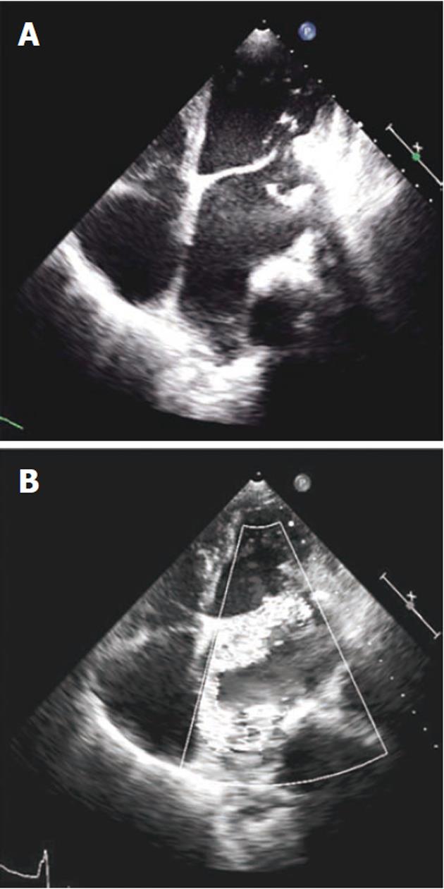
Figure 1 Echocardiography in apical 4 chamber view.
A: Calcified, thickened and retracted posterior mitral leaflet with adjacent mitral annular ring calcification; B: Color Doppler showing severe mitral regurgitation.
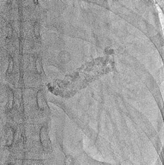
Figure 2 Fluoroscopy image in right anterior oblique 30° view showing severe mitral annular calcification.
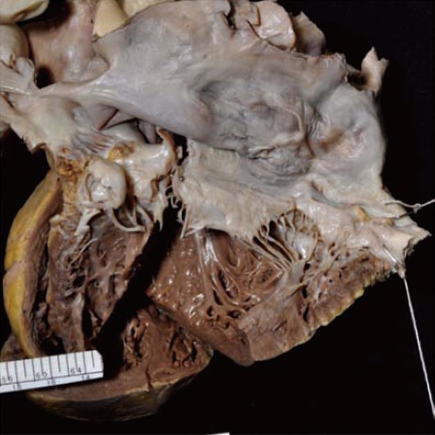
Figure 3 Gross photograph of the inflow tract of the left heart showing a grossly dilated left atrium and ventricle.
Both mitral leaflets show thickened and fused chordae. There is massive calcification of the posterior mitral annulus. Some of the calcified foci are visible as irregular nodules along the atrial surface of posterior leaflet.
- Citation: Vijayvergiya R, Vaiphei K, Rana SS. Severe mitral annular calcification in rheumatic heart disease: A rare presentation. World J Cardiol 2012; 4(3): 87-89
- URL: https://www.wjgnet.com/1949-8462/full/v4/i3/87.htm
- DOI: https://dx.doi.org/10.4330/wjc.v4.i3.87


