Copyright
©2012 Baishideng Publishing Group Co.
Figure 1 Coronary angiogram.
A: 90% diffuse stenosis of proximal left anterior descending artery (LAD) and 90% tubular stenosis of the distal left circumflex artery (LCx); B: No residual stenosis following proximal LAD and distal LCx stenting; C: At 15 mo of follow-up, showing patent LAD and LCx stents. There is new 70% bifurcation stenosis of the left main coronary arterywith osteal LAD stenosis. The LCx ostium is normal without any stenosis; D: No residual stenosis and normal LCx ostium following left main coronary artery crossover stenting.
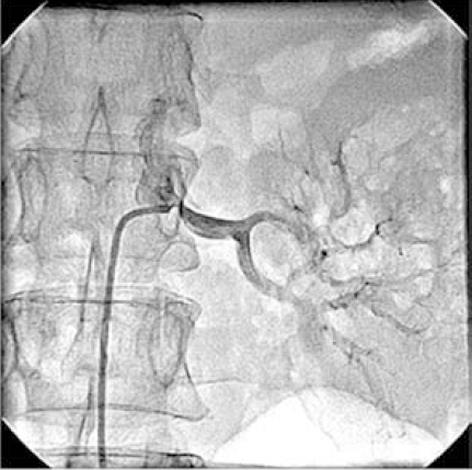
Figure 2 Left renal angiogram showing 75% osteal stenosis of renal artery.
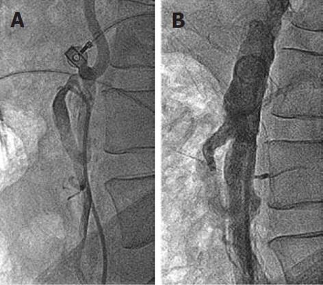
Figure 3 Inferior mesenteric artery angiogram.
A: 50% osteal stenosis of the inferior mesenteric artery (IMA); B: Post-IMA osteal stenting with a 7 mm x 18 mm balloon-expandable stent, at 15 mo follow-up, showing no residual IMA stenosis.
Figure 4 Abdominal aortogram.
A: Bilateral common iliac artery stenosis; B: No residual stenosis following left renal and aorto-bilateral iliac artery stenting. A big inferior mesenteric artery collateral “arch of Riolan” supplies the occluded superior mesenteric artery; C: At 30 mo follow-up, showing patent left renal and aorto-bilateral iliac stents.
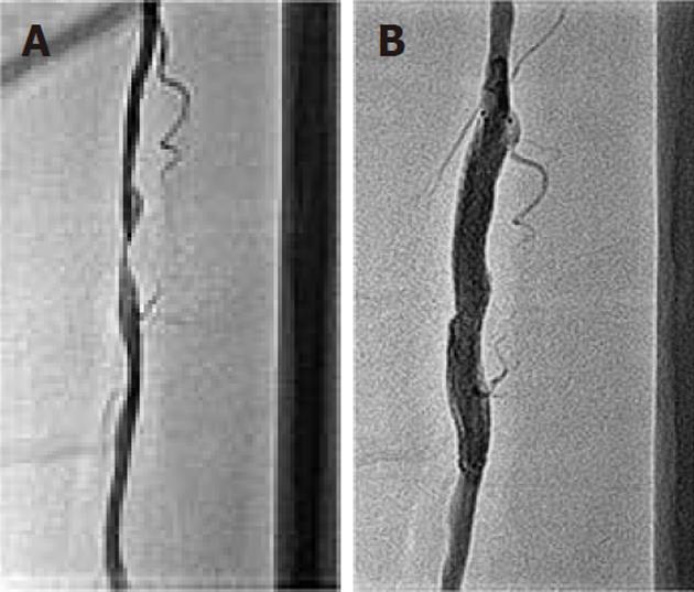
Figure 5 Left superficial femoral artery angiogram.
A: At 3 mo follow-up, showing 90% discrete stenosis in the mid part; B: Following 7 mm x 60 mm self-expanding stent deployment, there is no residual stenosis.
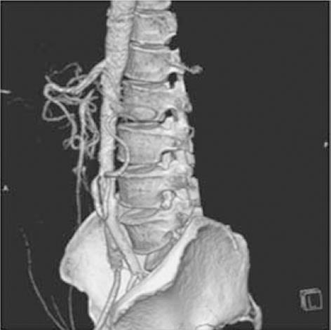
Figure 6 Computed tomography.
Image of the abdominal aorta at 9 mo follow-up, showing 90% stenosis of the inferior mesenteric artery (IMA) ostium. The totally occluded superior mesenteric artery at the ostium is filled retrogradely via the arch of Riolan from the IMA.
- Citation: Vijayvergiya R, Garg D, Sinha SK. Percutaneous panvascular intervention in an unusual case of extensive atherosclerotic disease. World J Cardiol 2012; 4(2): 48-53
- URL: https://www.wjgnet.com/1949-8462/full/v4/i2/48.htm
- DOI: https://dx.doi.org/10.4330/wjc.v4.i2.48


