Copyright
©2012 Baishideng Publishing Group Co.
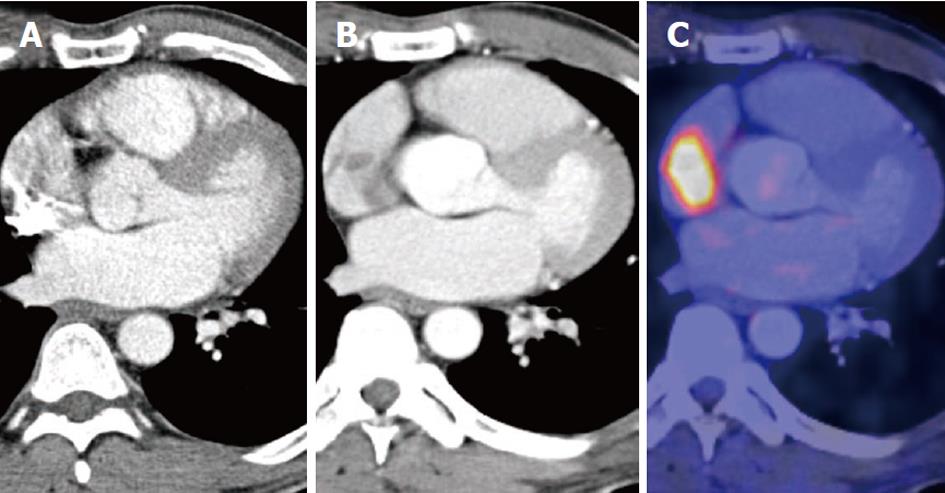
Figure 1 Computed tomography scan.
A: Initial computed tomography (CT) scan, with artifacts in the right atrium; B: CT of better quality; C: 18F-fluorodeoxyglucose positron emission tomography-CT with specific tracer accumulation.
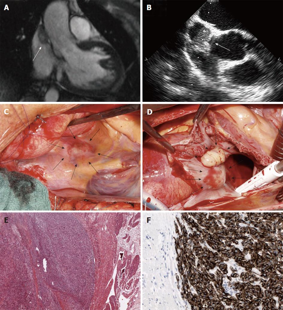
Figure 2 Tumor imaging and histology.
Magnetic resonance imaging (A) and transesophageal echocardiography (B) showing the tumor in the inflow of the right atrium. Intraoperative views of the superior vena cava from the outside (C) and the inside (D); Histologic examination reveals intracardiac metastasis of a malignant melanoma (E, F). Arrows indicate tumor localization.
- Citation: Krüger T, Heuschmid M, Kurth R, Stock UA, Wildhirt SM. Asymptomatic melanoma of the superior cavo-atrial junction: The challenge of imaging. World J Cardiol 2012; 4(1): 20-22
- URL: https://www.wjgnet.com/1949-8462/full/v4/i1/20.htm
- DOI: https://dx.doi.org/10.4330/wjc.v4.i1.20


