Copyright
©2011 Baishideng Publishing Group Co.
World J Cardiol. Sep 26, 2011; 3(9): 311-314
Published online Sep 26, 2011. doi: 10.4330/wjc.v3.i9.311
Published online Sep 26, 2011. doi: 10.4330/wjc.v3.i9.311
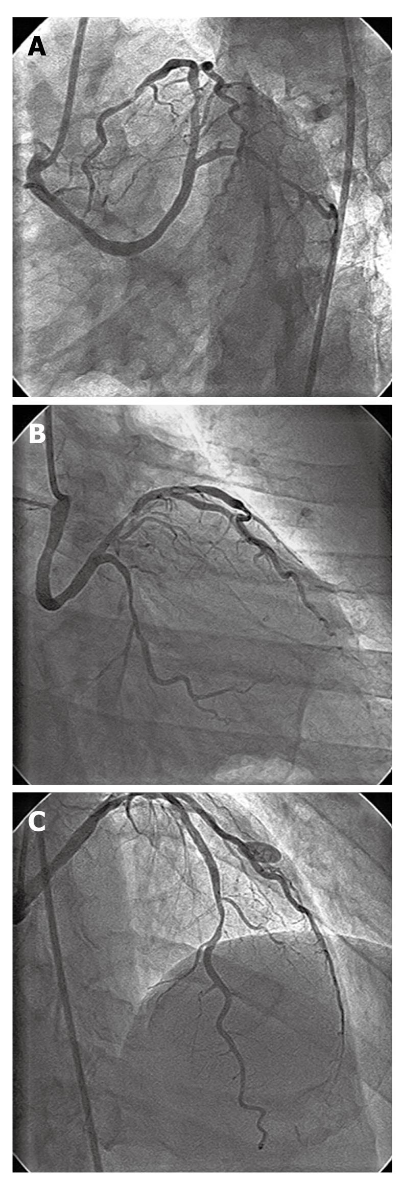
Figure 1 Left coronary angiogram after injection from right coronary sinus.
A: Separate ostial origin of anomalous left main coronary artery from right coronary sinus, having retro-aortic course; B: Proximal left anterior descending (LAD) artery having type B 70% tubular stenosis, proximal left circumflex artery having 50% diffuse stenosis; C: Distal LAD artery having type B 70% tubular stenosis.
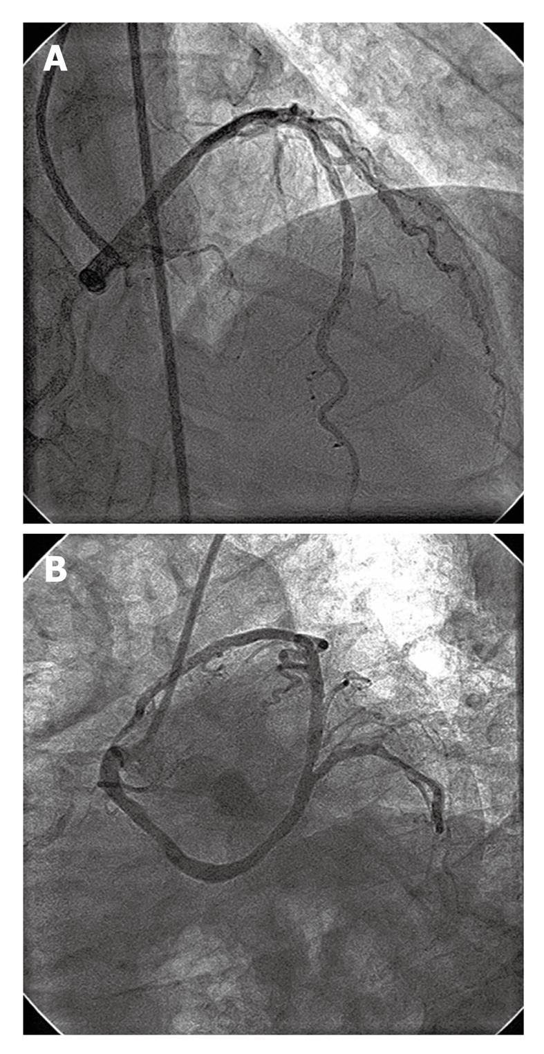
Figure 2 Left coronary angiogram following coronary stenting of left anterior descending artery.
A: Distal left anterior descending (LAD) lesion stented with 2.5 mm × 23 mm sirolimus-eluting stent; B: Proximal LAD lesion stented with 3 mm × 23 mm sirolimus-eluting stent.
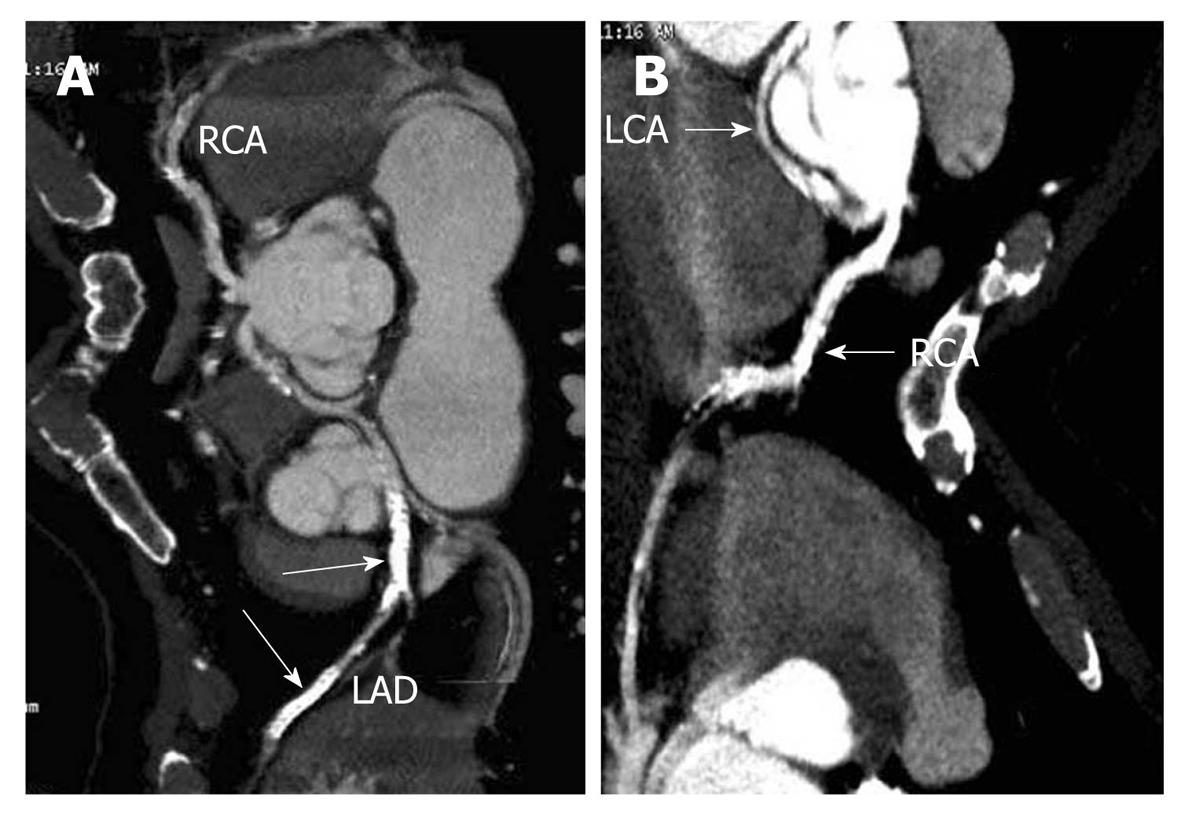
Figure 3 64-slice multi-detector computed tomography image of coronary arteries.
A: Computed tomography (CT) image showing separate ostia of both right and left main coronary artery. The left anterior descending stents are marked with white arrow; B: CT image showing retro-aortic course of left main coronary artery. RCA: Right coronary artery; LCA: Left coronary artery; LAD: Left anterior descending.
- Citation: Vijayvergiya R, Grover A, Singhal M. Percutaneous revascularization in a patient with anomalous origin of left main coronary artery. World J Cardiol 2011; 3(9): 311-314
- URL: https://www.wjgnet.com/1949-8462/full/v3/i9/311.htm
- DOI: https://dx.doi.org/10.4330/wjc.v3.i9.311


