Copyright
©2011 Baishideng Publishing Group Co.
World J Cardiol. May 26, 2011; 3(5): 165-168
Published online May 26, 2011. doi: 10.4330/wjc.v3.i5.165
Published online May 26, 2011. doi: 10.4330/wjc.v3.i5.165
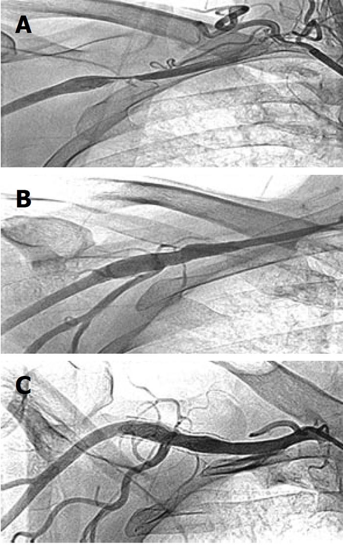
Figure 1 Peripheral angiogram of axillary artery stenosis and its endovascular treatment in case 1.
A: Angiogram showing 90% short segment stenosis of proximal part of right axillary artery; B: Brisk flow with no residual stenosis of axillary artery following a 8 mm × 40 mm self expanding nitinol stent deployments; C: At 10 mo of follow-up, patent axillary stent with no in-stent restenosis and brisk flow across it.
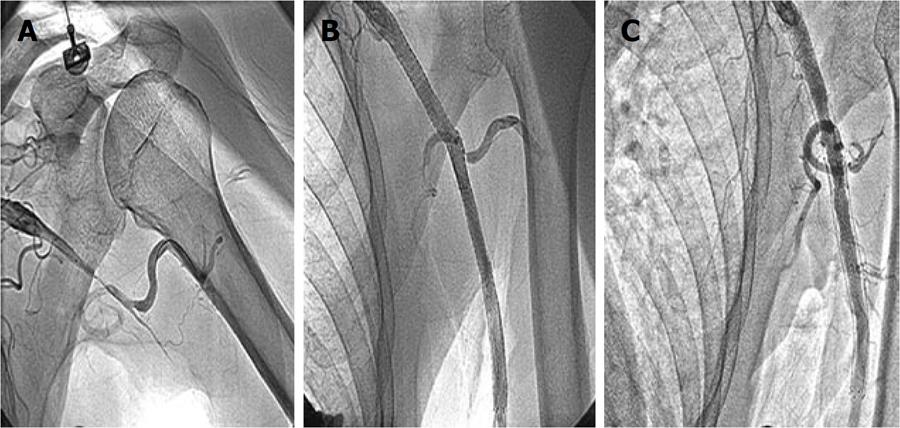
Figure 2 Peripheral angiogram of axillary artery stenosis and its endovascular treatment in case 2.
A: Total occlusion of distal part of left axillary and brachial artery; B: Brisk flow across the axillary-brachial segment following two 8 mm × 80 mm, 8 mm × 60 mm self expanding nitinol stent deployment; C: At 5 mo of follow-up, patent axillary stent and brisk flow across it.
- Citation: Vijayvergiya R, Yadav M, Grover A. Percutaneous endovascular management of atherosclerotic axillary artery stenosis: Report of 2 cases and review of literature. World J Cardiol 2011; 3(5): 165-168
- URL: https://www.wjgnet.com/1949-8462/full/v3/i5/165.htm
- DOI: https://dx.doi.org/10.4330/wjc.v3.i5.165


