Copyright
©2011 Baishideng Publishing Group Co.
World J Cardiol. May 26, 2011; 3(5): 127-134
Published online May 26, 2011. doi: 10.4330/wjc.v3.i5.127
Published online May 26, 2011. doi: 10.4330/wjc.v3.i5.127
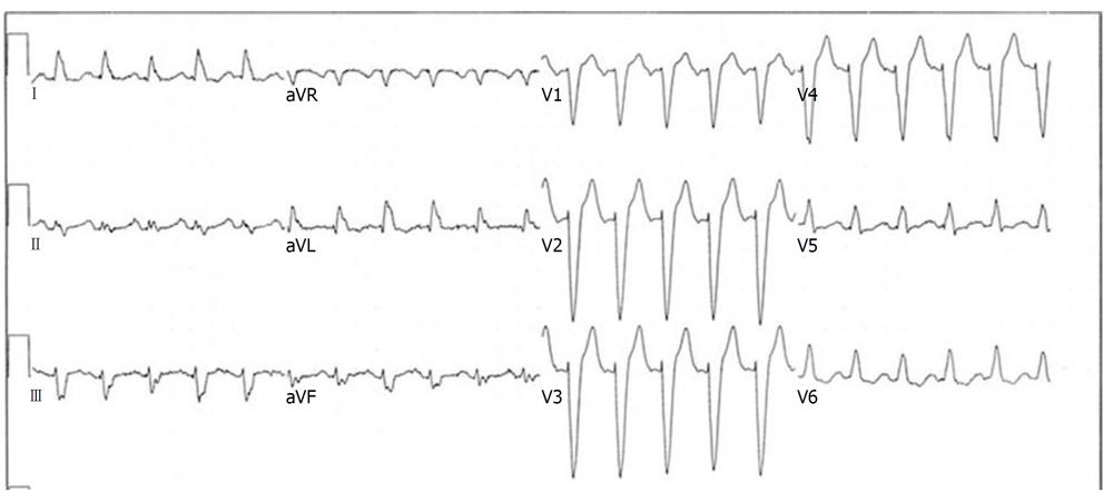
Figure 1 Electrocardiogram example of typical left bundle branch block pattern in a patient with sinus tachycardia.
Lead V1 demonstrates an rS complex, while there are monophasic, notched R waves in leads I, aVL, V5 and V6. Q waves are absent in these leads.
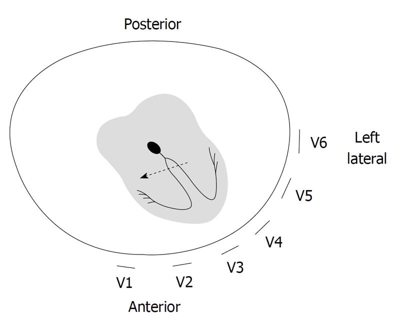
Figure 2 Mechanism of left bundle branch block-like electrocardiogram morphology.
Electrical activation arises from outside the specialized conduction system and travels rightward before activating the left ventricle. This results in a Q wave in the left lateral electrocardiogram leads. Adapted from[8], with permission.
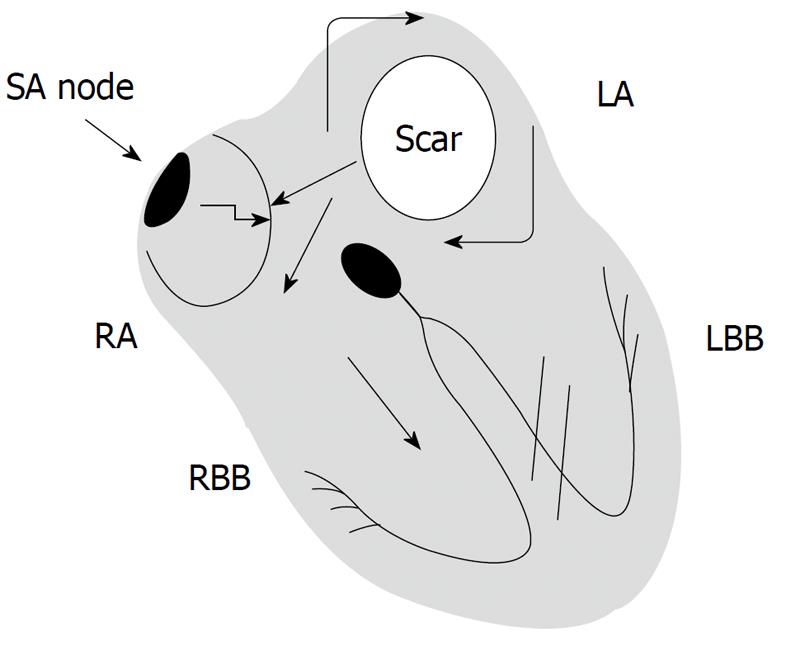
Figure 3 Atrial tachycardia can result in functional left bundle branch block.
A reentrant circuit in the left atrium results in tachycardia that rapidly conducts to the ventricle via the atrioventricular node. Rate-dependent conduction block occurs in the left bundle branch. Adapted from[10], by permission of Oxford University Press, Inc. LBB: Left bundle branch; RBB: Right bundle branch.
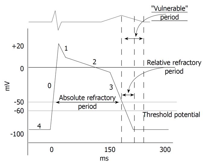
Figure 4 Typical action potential from within the His-Purkinje system.
If this potential was from the left bundle branch, impulses that occur (encroach upon) the absolute refractory period will not excite the tissue. A new action potential will not occur and (assuming it is not also refractory) conduction will occur solely through the right bundle branch. Adapted from[11], with permission.
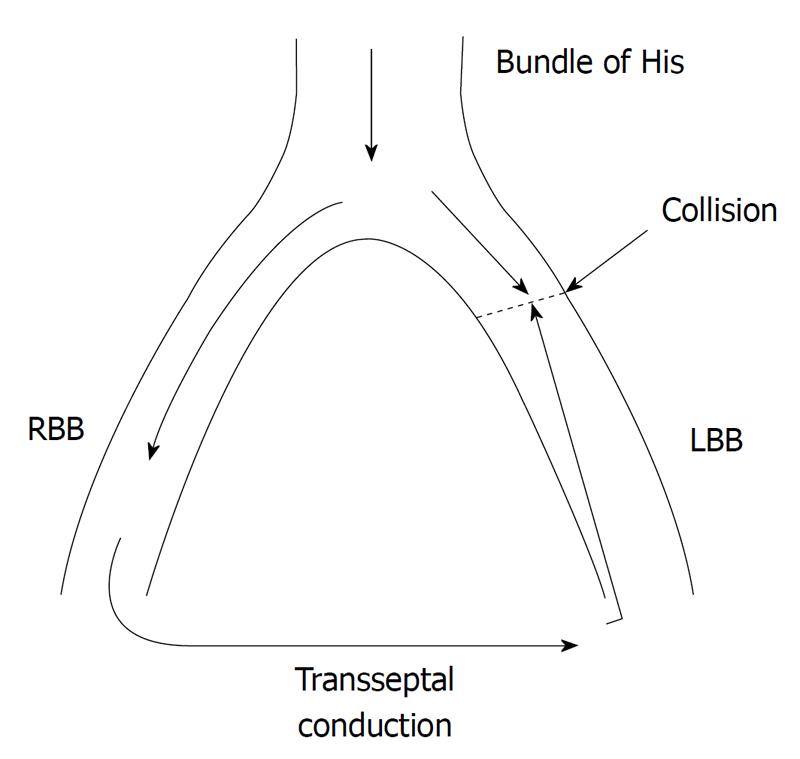
Figure 5 Mechanism of linking in functional left bundle branch block aberrance.
Repetitive transseptal retrograde concealed penetration from impulses conducting antegrade via the right bundle perpetuates local refractoriness or results in repetitive impulse collision. LBB: Left bundle branch; RBB: Right bundle branch.
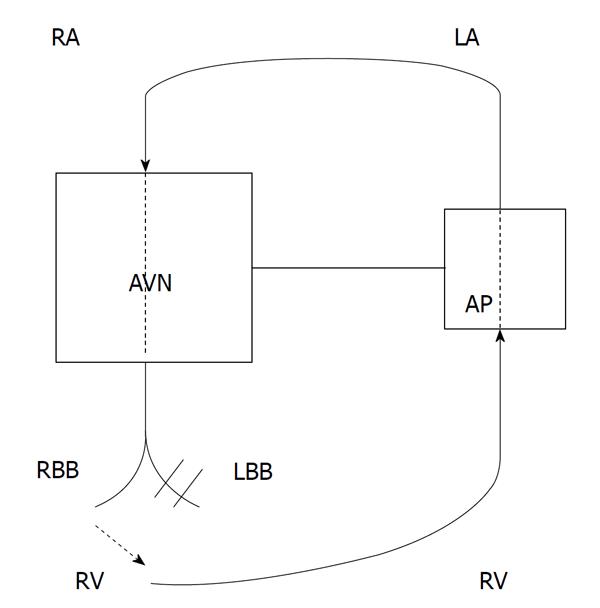
Figure 6 Illustration of orthodromic AV reentry tachycardia resulting in a wide QRS tachycardia with left bundle branch block morphology.
A left sided accessory pathway is present. During tachycardia, antegrade conduction is via the atrioventricular node and right bundle branch (RBB), and retrograde conduction is via the accessory pathway. Adapted from[15], with permission. LBB: Left bundle branch.
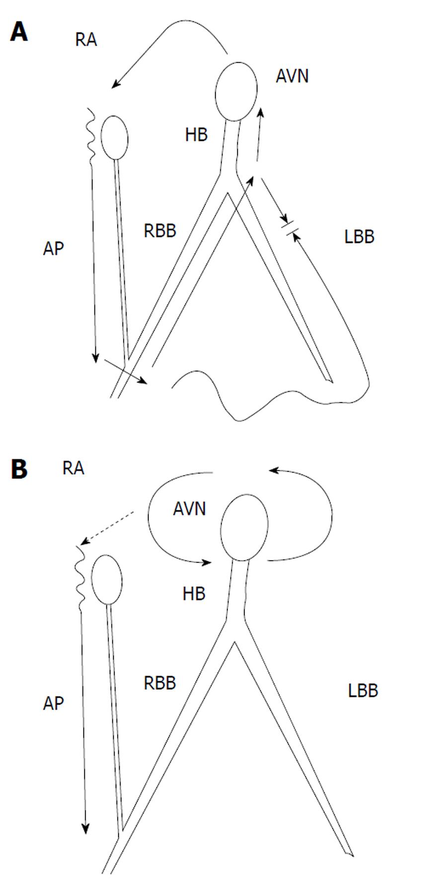
Figure 7 Pre-excitation via atriofascicular (“Mahaim”) bypass tracts.
A: Antidromic AV reentry tachycardia. The tachycardia circuit conducts antegrade down the Mahaim fiber and inserts into the distal right bundle branch (RBB). It propagates retrograde back to the atrium via the more proximal portion of the RBB; B: Atrioventricular nodal reentrant tachycardia with a “bystander” Mahaim fiber present. The bypass tract is not part of the tachycardia circuit, but contributes to ventricular activation. A wide QRS complex will be present with a left bundle branch block configuration, but ablation of the bypass tract will not eliminate the tachycardia. Adapted from[18], with permission. LBB: Left bundle branch.
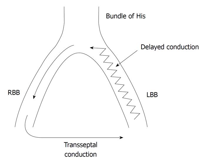
Figure 8 Mechanism of bundle branch reentrant ventricular tachycardia.
The right bundle branch (RBB) is the antegrade limb of the circuit, with retrograde conduction via the slowly conducting left bundle branch. This allows the RBB to recover and be capable of reactivation, thereby perpetuating the reentrant circuit. LBB: Left bundle branch.
- Citation: Neiger JS, Trohman RG. Differential diagnosis of tachycardia with a typical left bundle branch block morphology. World J Cardiol 2011; 3(5): 127-134
- URL: https://www.wjgnet.com/1949-8462/full/v3/i5/127.htm
- DOI: https://dx.doi.org/10.4330/wjc.v3.i5.127


