Copyright
©2011 Baishideng Publishing Group Co.
World J Cardiol. Mar 26, 2011; 3(3): 98-100
Published online Mar 26, 2011. doi: 10.4330/wjc.v3.i3.98
Published online Mar 26, 2011. doi: 10.4330/wjc.v3.i3.98
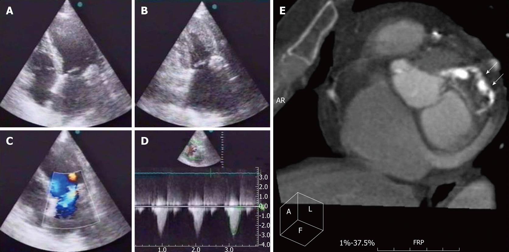
Figure 1 Pre-discharge transthoracic echocardiogram.
Apical 4C (panel A) and 2C (panel B) showing a 2.3 cm × 2.0 cm round echodense mass at the posterior side of the mitral annulus, grade II mitral regurgitation (panel C) and moderate aortic stenosis (peak gradient 33 and medium 21 mmHg) (panel D); E: Cardiac assessment with 16-slice multidetector row computed tomography (Philips Brilliance 16 using electrocardiographic triggering, endovenous contrast enhancement and multiplanar and volumetric reconstruction), a peripherally calcified, round 2.5 cm × 3.0 cm mass with decreased signal on the inside can be seen in the posterior part of the mitral annulus (arrows).
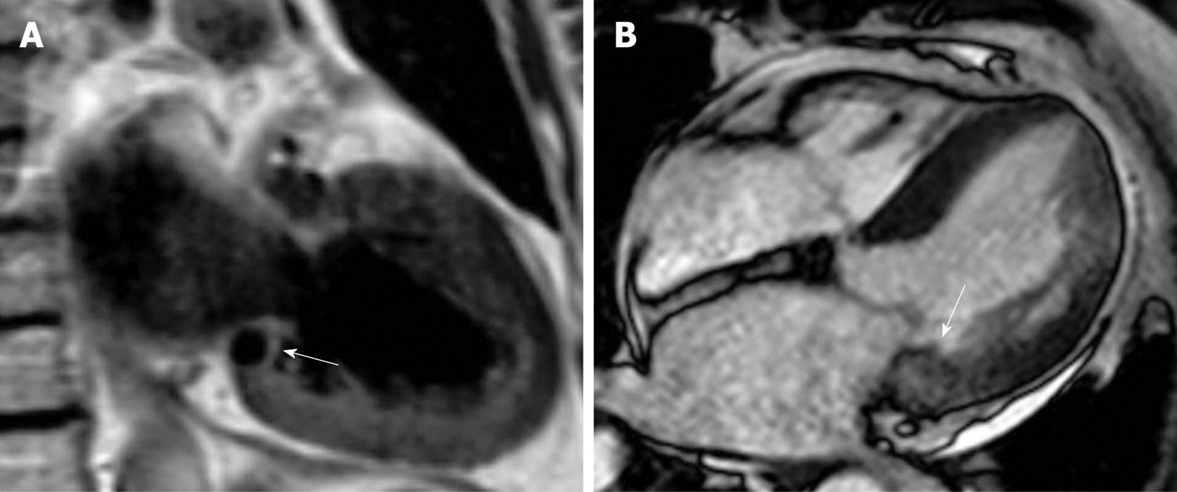
Figure 2 Cardiac magnetic resonance images (Phillips 1.
5 Tesla MRI scanner). A: A well defined dark mass (arrow) is near the posterior mitral valve leaflet in a T1 weighted sequence, suggestive of calcification; B: In steady state free precession images, the mass (arrow) appears only slightly darker than the normal myocardium, with a well defined intramyocardial border.
- Citation: Providência R, Botelho A, Mota P, Catarino R, Leitão-Marques A. Cardiac mass in a patient with colorectal cancer: “Not all that glitters is gold!”. World J Cardiol 2011; 3(3): 98-100
- URL: https://www.wjgnet.com/1949-8462/full/v3/i3/98.htm
- DOI: https://dx.doi.org/10.4330/wjc.v3.i3.98


