Copyright
©2011 Baishideng Publishing Group Co.
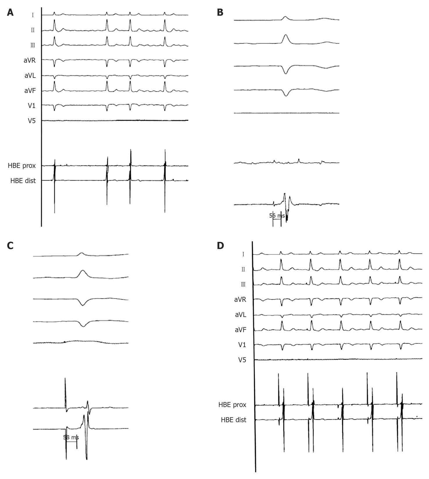
Figure 1 Spontaneous and Hisian paced ECGs.
A: Surface electrocardiogram (ECG; peripheral derivations and V1-V5) and endocardial electrogram (EGM; His bundle site) during chronic atrial fibrillation: the QRS is narrow (90 ms) (registration speed 25 mm/s); B: His bundle registration: HV 56 ms (registration speed 50 mm/s); C: Spike-V during direct Hisian pacing equal to the basal HV (58 ms) (registration speed 100 mm/s); D: Surface ECG (peripheral derivations and V1-V5) and endocardial EGM (His bundle site) during direct Hisian pacing: the QRS is equal to the native QRS (registration speed 25 mm/s).
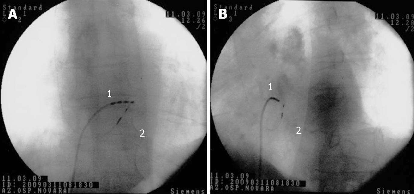
Figure 2 His bundle pacing.
Antero-posterior (A) and left anterior oblique (B) fluoroscopic projections showing the screw-in lead (Select Secure, Medtronic) position during the procedure for a direct His bundle pacing; 1 = quadripolar Hisian mapping catheter; 2 = screw-in bipolar lead positioned in close proximity to the His bundle.
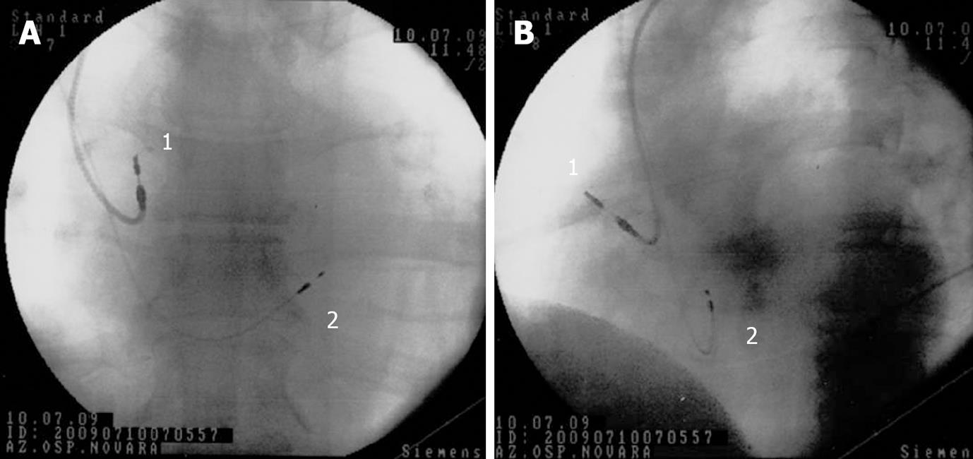
Figure 3 Para-Hisian pacing.
Antero-posterior (A) and left anterior oblique (B) fluoroscopic projections showing lead positions after dual-chamber atrio-ventricular cardiac para-Hisian pacing. 1 = conventional screw-in atrial lead, placed into the right atrium; 2 = screw-in bipolar lead (Select Secure, Medtronic) positioned near the His bundle.
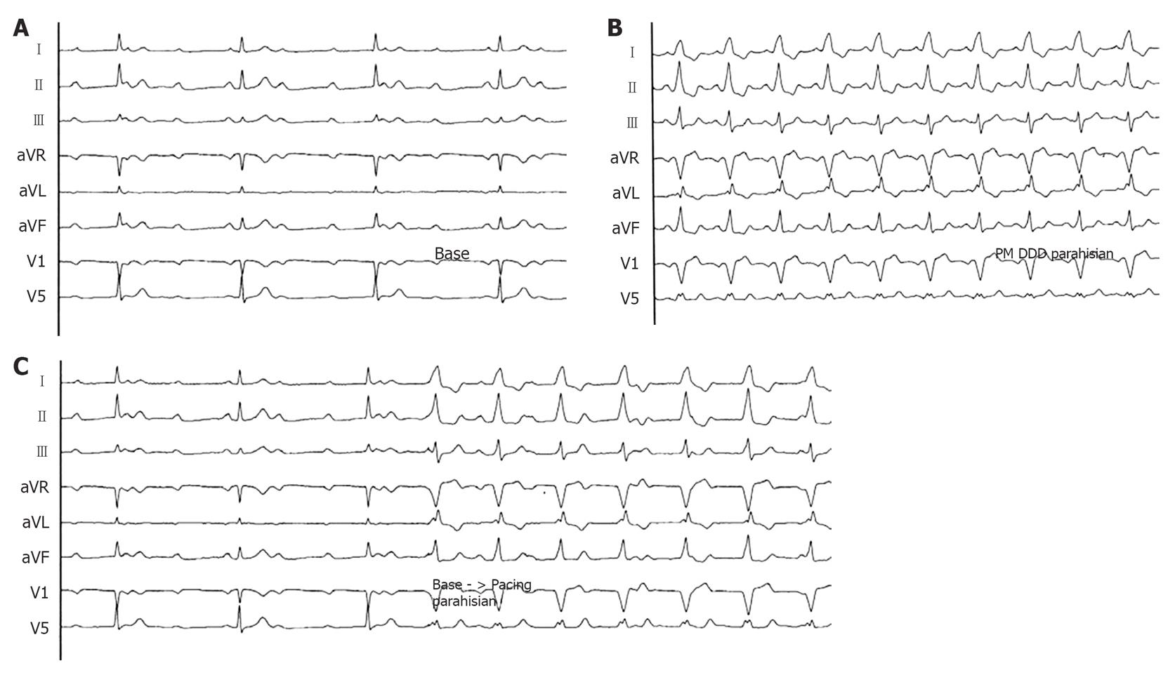
Figure 4 Spontaneous and para-Hisian paced ECGs.
A: Surface ECG (peripheral derivations and V1-V5) during complete AV block with narrow QRS (90 ms) (registration speed 25 mm/s); B: Surface ECG (peripheral derivations and V1-V5) during para-Hisian pacing: the QRS is larger respect to the native QRS (registration speed 25 mm/s); C: Passage from to the native QRS to the para-Hisian paced QRS: the electrical axis is exactly the same (registration speed 25 mm/s). DDD: Dual-chamber atrio-ventricular cardiac pacing.
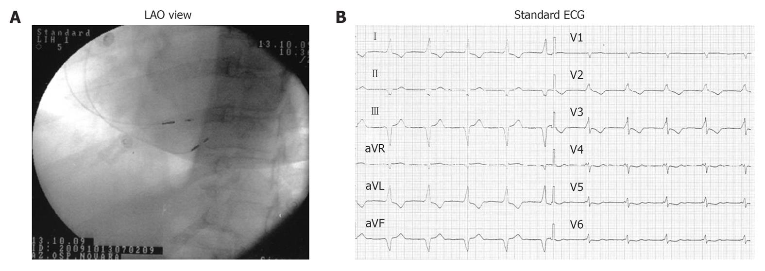
Figure 5 Atrial septal and para-Hisian dual-chamber atrio-ventricular cardiac pacing.
A: left anterior oblique (LAO) fluoroscopic projections showing atrial septal lead and para-Hisian ventricular lead positions; B: 12-lead surface ECG during dual-chamber atrio-ventricular cardiac septal pacing (atrial septal lead and para-Hisian ventricular lead).
- Citation: Occhetta E, Bortnik M, Marino P. Future easy and physiological cardiac pacing. World J Cardiol 2011; 3(1): 32-39
- URL: https://www.wjgnet.com/1949-8462/full/v3/i1/32.htm
- DOI: https://dx.doi.org/10.4330/wjc.v3.i1.32


