Copyright
©2010 Baishideng Publishing Group Co.
World J Cardiol. Aug 26, 2010; 2(8): 223-232
Published online Aug 26, 2010. doi: 10.4330/wjc.v2.i8.223
Published online Aug 26, 2010. doi: 10.4330/wjc.v2.i8.223
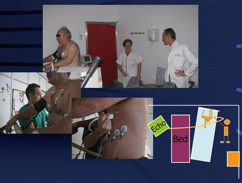
Figure 1 Exercise echocardiography.
When the patient is exhausted or termination criteria appear (symptoms, significative ST changes, decrease or increase in blood pressure, etc.), the observer acquires images by placing the transducer in the cardiac apex, then in the parasternal region. Note the placement of the table, the treadmill and the echocardiography machine for feasible imaging evaluation at peak and post-exercise. The left lateral handlebar of the treadmill has been removed to allow for rapid post-exercise positioning of the patient on the table.
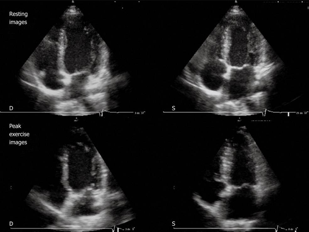
Figure 2 Resting echocardiography (top) and peak exercise echocardiography (bottom).
Four-chamber apical view (diastolic frames on the left, systolic frames on the right) in a patient with normal results. Note the left ventricular (LV) cavity dimensions decrease with exercise and an increase in LV ejection fraction. D: Diastolic; S: Systolic.
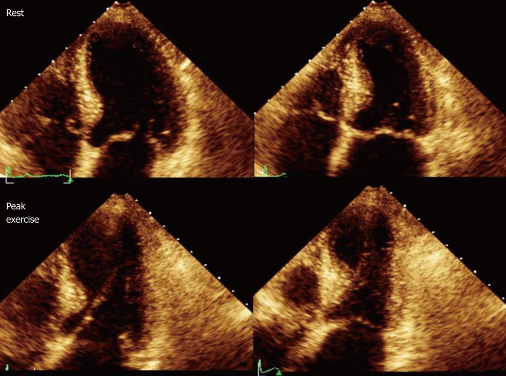
Figure 3 Resting echocardiography (top) and peak exercise echocardiography (bottom).
Four-chamber apical view (diastolic frames on the left, systolic frames on the right) in a patient with significant coronary stenoses in the left anterior descending artery (99%). At rest, wall motion is normal, whereas during exercise a septoapical dyssynergia is observed with the typical 8-shaped left ventricular.
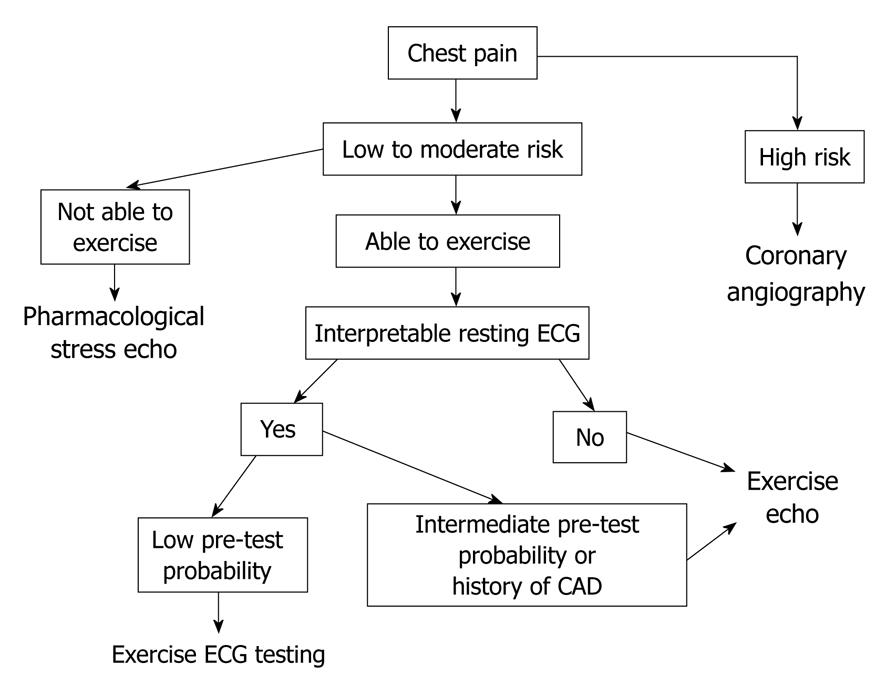
Figure 4 Algorithm used in our institution for patients with chest pain.
CAD: Coronary artery disease; ECG: Electrocardiography.
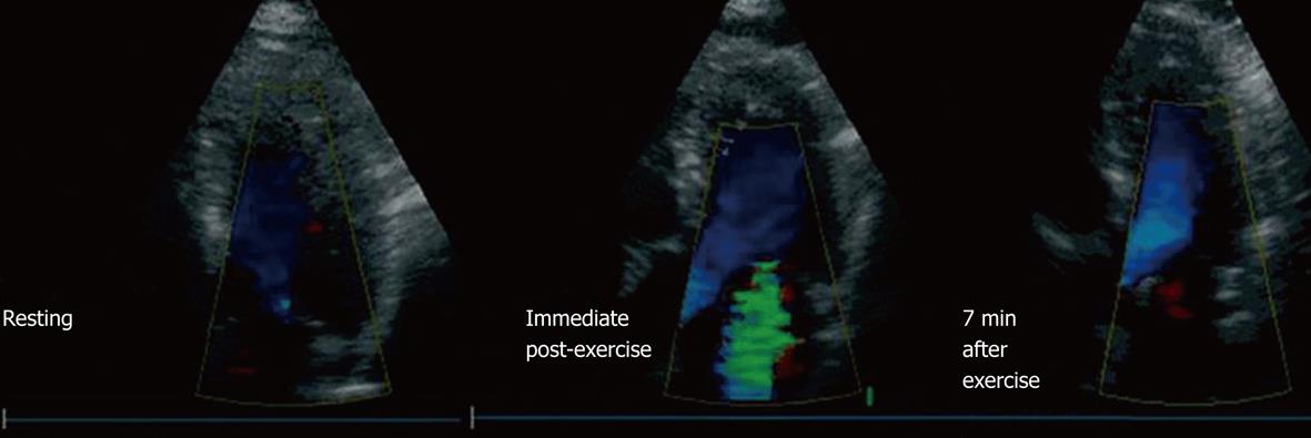
Figure 5 Example of a patient with mild ventricular dysfunction (resting left ventricle ejection fraction 49%, exercise left ventricle ejection fraction 46%) who developed severe mitral regurgitation (MR) during exercise.
This patient had no MR at rest (left), severe MR developed in the immediate post-exercise period (center), which did not completely disappear until 7 min after exercise (right) (From Peteiro et al[32]).
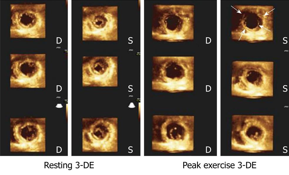
Figure 6 Cropped views obtained from a left ventricle full-volume during 3-dimensional exercise echocardiography in resting conditions (left panel) and during peak exercise (right panel) in a patient with severe 3-vessel disease.
Note exercise-induced akinesia and dilation in the short axis apical views (arrows), as well as hypokinesia and dilation in the short-axis view at the papillary muscles level. 3-DE: Three-dimensional exercise; D: Diastolic; S: Systolic.
- Citation: Peteiro J, Bouzas-Mosquera A. Exercise echocardiography. World J Cardiol 2010; 2(8): 223-232
- URL: https://www.wjgnet.com/1949-8462/full/v2/i8/223.htm
- DOI: https://dx.doi.org/10.4330/wjc.v2.i8.223


