Copyright
©2010 Baishideng Publishing Group Co.
World J Cardiol. Oct 26, 2010; 2(10): 333-343
Published online Oct 26, 2010. doi: 10.4330/wjc.v2.i10.333
Published online Oct 26, 2010. doi: 10.4330/wjc.v2.i10.333
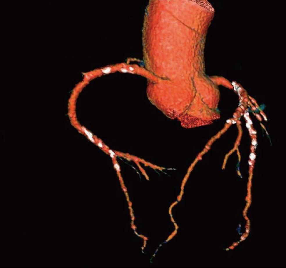
Figure 1 Three-dimensional volume rendering of the left and right coronary arteries and their branches acquired with 64-slice computed tomography angiography in a 61-year-old diagnosed with coronary artery disease.
Extensive calcification is present in the coronary artery wall which is shown as the white dots.
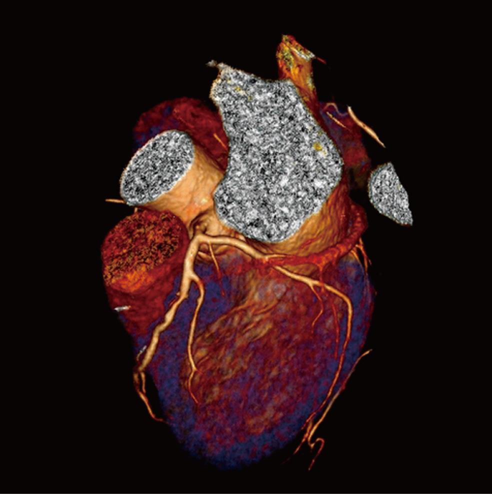
Figure 2 Three-dimensional volume rendering of the left coronary artery is acquired with dual-source computed tomography angiography in a 47-year-old woman suspected of coronary artery disease.
Left anterior descending and left circumflex are clearly demonstrated without any sign of lumen stenosis or calcification.
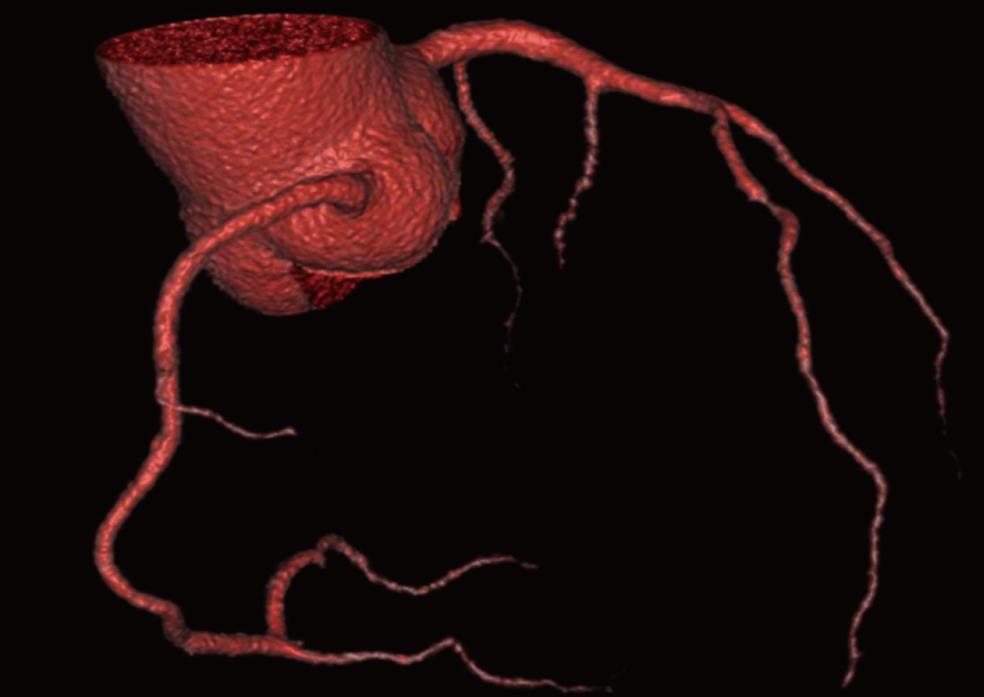
Figure 3 Three-dimensional volume rendering of the coronary arteries and side branches are clearly visualized with use of 320-slice computed tomography angiography in a 58-year-old man presenting with chest pain.
Volumetric data were acquired within a single heartbeat with excellent image quality.
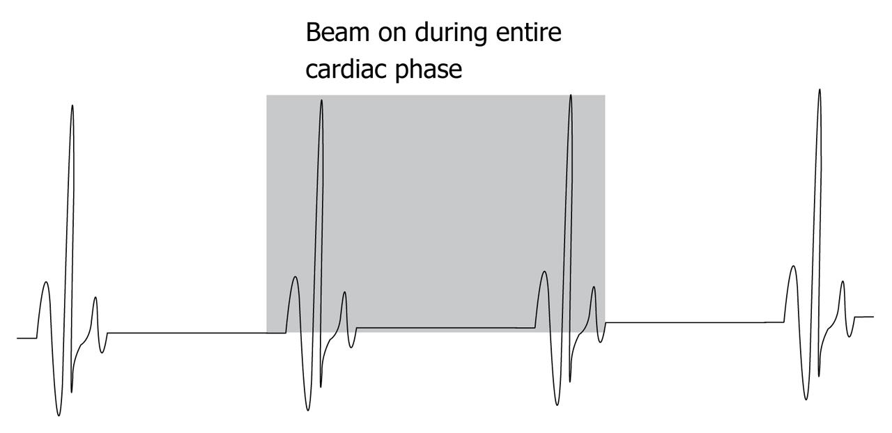
Figure 4 Diagram showing retrospective electrocardiogram-gating without tube current modulation in multislice computed tomography coronary angiography.
An X-ray beam is turned on during the entire cardiac cycle without adjusting the tube current.
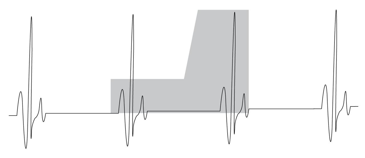
Figure 5 Diagram showing retrospective electrocardiogram-gating with the use of tube current modulation in multislice computed tomography coronary angiography.
Normal tube current is applied only during the image reconstruction phase (late diastolic phase), while the tube current is reduced significantly during the systolic phase.
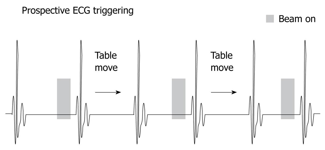
Figure 6 The diagram shows prospective electrocardiogram-triggering with X-ray beam on during a portion of the cardiac cycle, while in the remaining cardiac phase, the X-ray beam is turned off.
Figure 7 Characterization of coronary plaques.
RCA-right coronary artery. A: Curved planar reformatted image acquired with 64-slice computed tomography angiography shows a non-calcified plaque at the mid-segment of the right coronary artery in a 57-year-old man suspected of coronary artery disease; B: Calcified plaques (arrows) are found at the proximal and middle segments of the right coronary artery; C: Mixed type plaques are shown in the left anterior descending (LAD) artery in a 59-year-old man suspected of coronary artery disease; the long arrow indicates the calcified plaque, while the short arrows indicate non-calcified components.
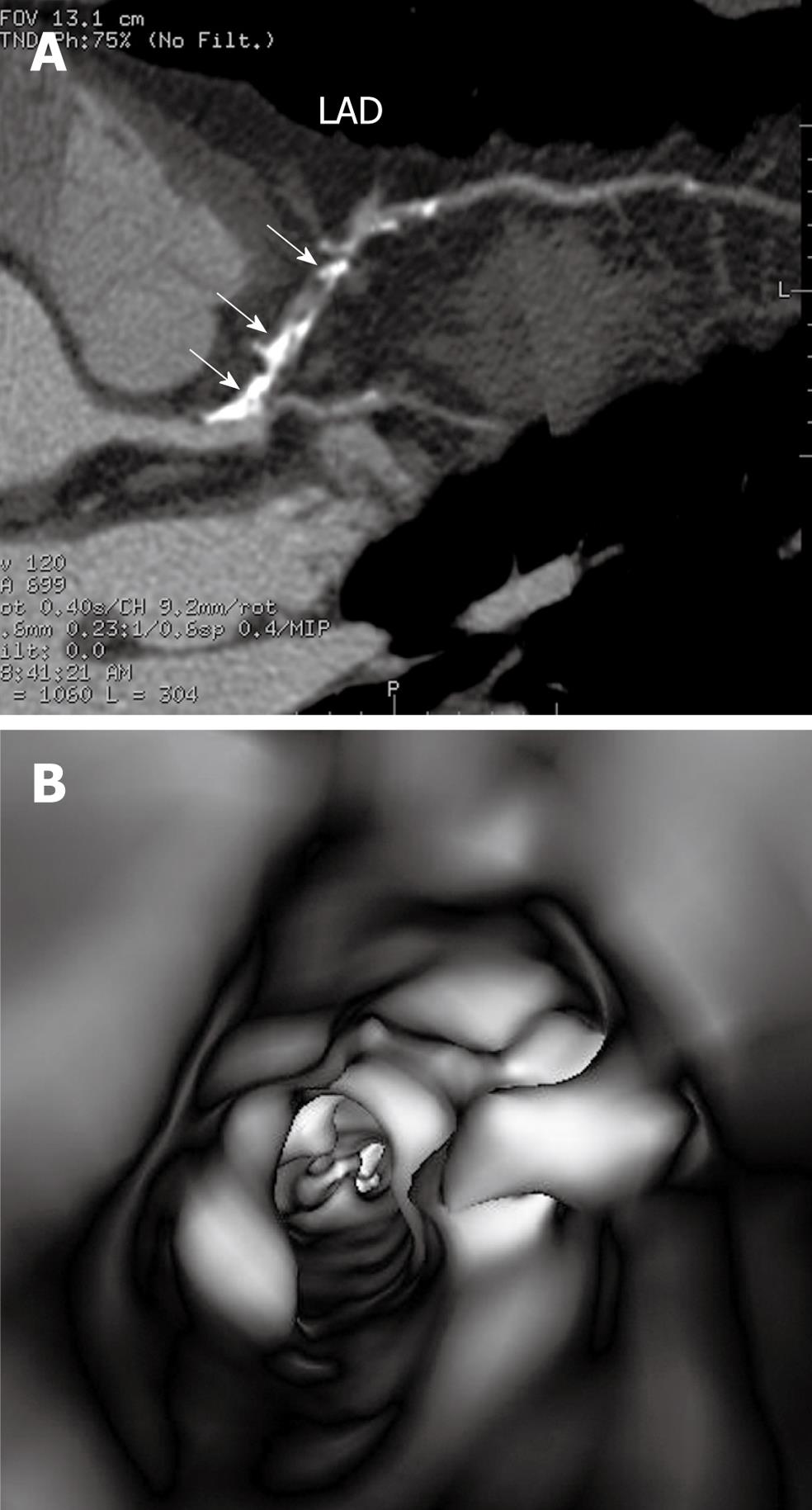
Figure 8 Virtual endoscopy visualization of coronary plaques.
A: Extensive calcified plaques in the left anterior descending (LAD) are observed on curved planar reformatted view with more than 70% luminal stenosis in a 52-year-old man with chest pain; B: Corresponding virtual intravascular endoscopy visualization demonstrates irregular intraluminal appearance with significant stenosis of the coronary artery.
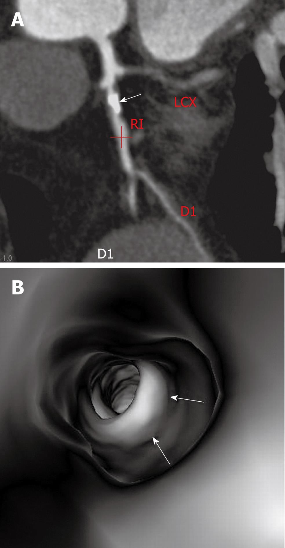
Figure 9 Virtual endoscopy confirmation of plaque stenosis.
A: A calcified plaque (arrow) is present in the left anterior descending with more than 90% lumen stenosis in a 51-year-old man presenting with symptoms of chest pain; B: Corresponding virtual intravascular endoscopy shows intraluminal protrusion caused by the plaque, but the luminal stenosis is less than 70%; arrows indicate the intraluminal appearance of calcified plaque. RI: Ramus intermedius; LCX: left circumflex; D1: Diagonal branch.
- Citation: Sun Z. Multislice CT angiography in coronary artery disease: Technical developments, radiation dose and diagnostic value. World J Cardiol 2010; 2(10): 333-343
- URL: https://www.wjgnet.com/1949-8462/full/v2/i10/333.htm
- DOI: https://dx.doi.org/10.4330/wjc.v2.i10.333


