Copyright
©2010 Baishideng Publishing Group Co.
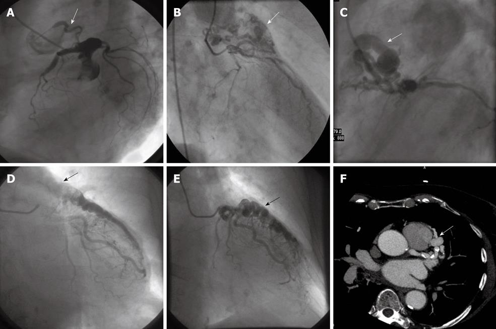
Figure 1 Different morphologic appearances of coronary artery-pulmonary artery fistulas (arrows).
A: Left lateral view of coronary angiogram demonstrating a fistula with a single origin, tortuous pathway and single termination (arrow); B: Right anterior oblique (RAO) view showing a fistula with multiple origin and outflow associated with a plexiform pathway (arrow). Shallow filling of the coronary arteries is visible; C: Left lateral projection illustrating a fistula with multiple origin and termination associated with aneurysmal formation (arrow), and total occlusion of the left anterior descending coronary artery (LAD) is appreciated; D and E: Sequential frames in RAO projection depicting a huge tortuous LAD fistulating into the pulmonary artery (arrows); F: Multislice cardiac gated computed tomography scan demonstrating the fistula running from the LAD into the pulmonary artery (arrow), and the fistula is connected to the pulmonary artery at the anterior side.
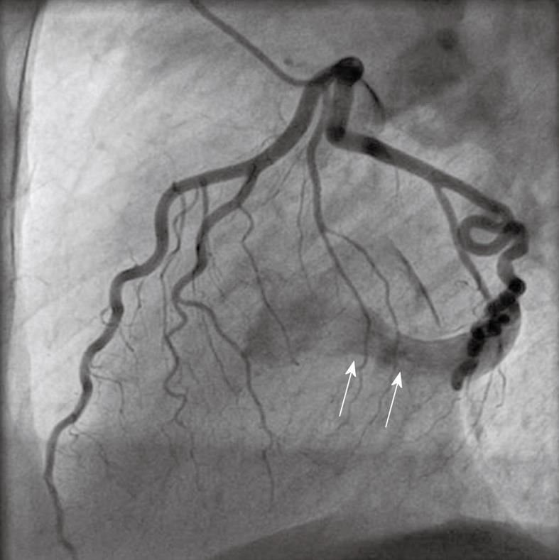
Figure 2 A fistula originating from the circumflex coronary artery and terminating into the coronary sinus (arrows).
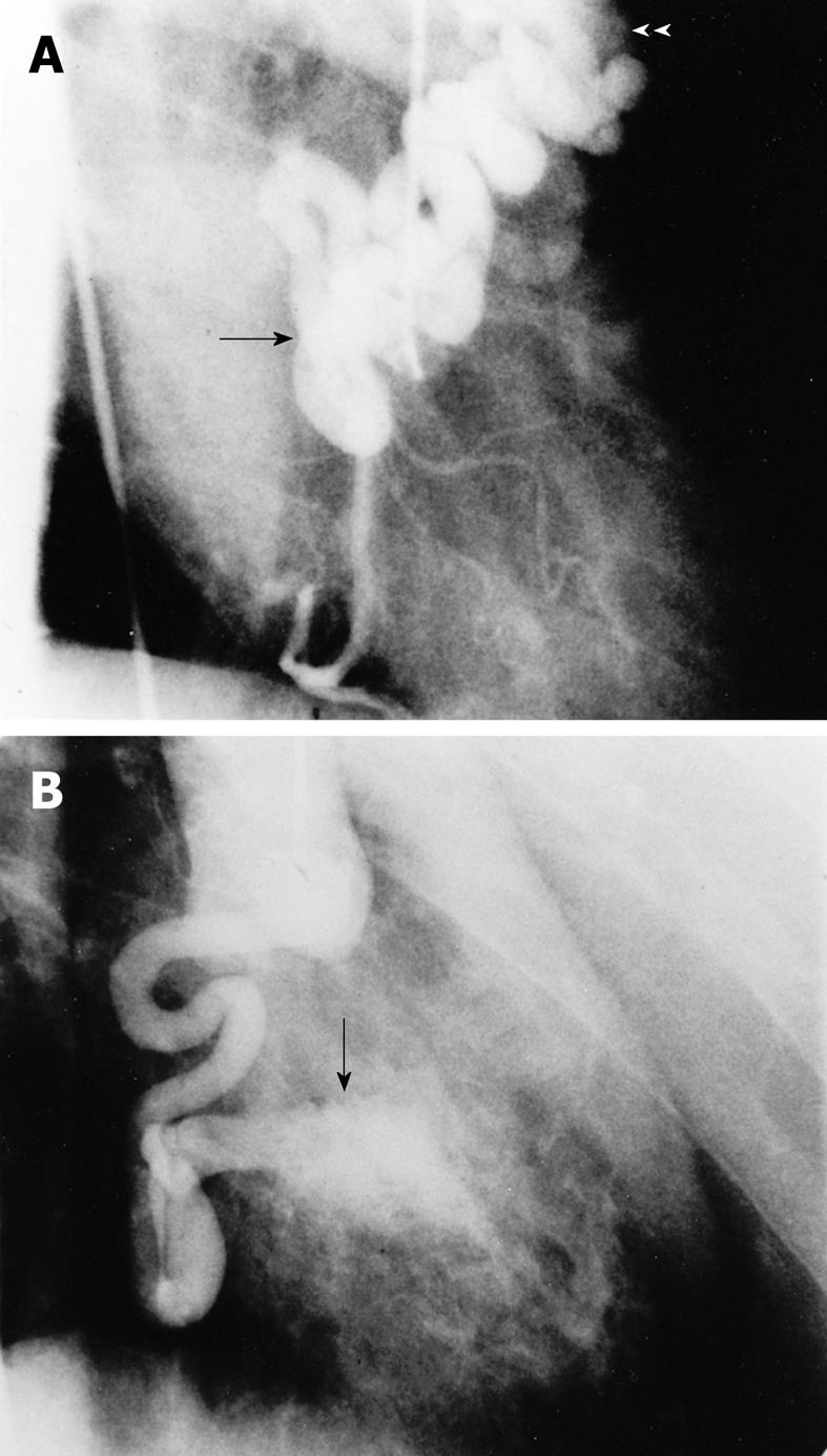
Figure 3 Different morphologic appearances of a dilated right coronary artery terminating into the pulmonary artery and the left ventricle (arrows).
A: RAO projection of a fistula originating from the proximal segment (arrow) of the right coronary artery and terminating into the pulmonary artery (arrowheads); B: A fistula from the distal segment (arrow) ending to the left ventricle.
- Citation: Said SA. Congenital solitary coronary artery fistulas characterized by their drainage sites. World J Cardiol 2010; 2(1): 6-12
- URL: https://www.wjgnet.com/1949-8462/full/v2/i1/6.htm
- DOI: https://dx.doi.org/10.4330/wjc.v2.i1.6


