Copyright
©The Author(s) 2025.
World J Cardiol. Jul 26, 2025; 17(7): 108745
Published online Jul 26, 2025. doi: 10.4330/wjc.v17.i7.108745
Published online Jul 26, 2025. doi: 10.4330/wjc.v17.i7.108745
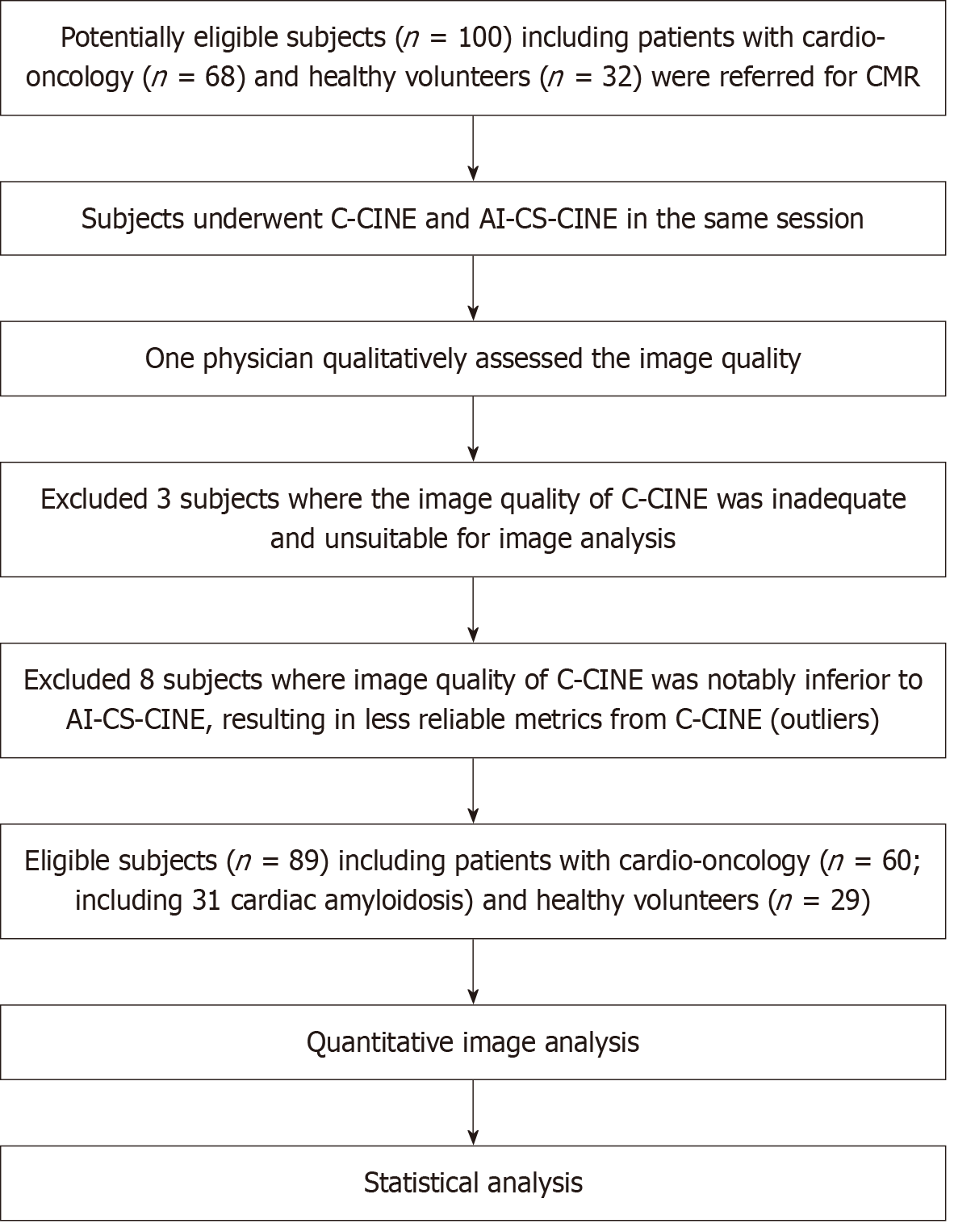
Figure 1 The flowchart shows exclusion criteria and the resulting cardio-oncology and healthy participants eligible and included in this study.
C-CINE: Conventional CINE; AI-CS-CINE: Artificial-intelligence-assisted compressed sensing CINE; CMR: Cardiac magnetic resonance.
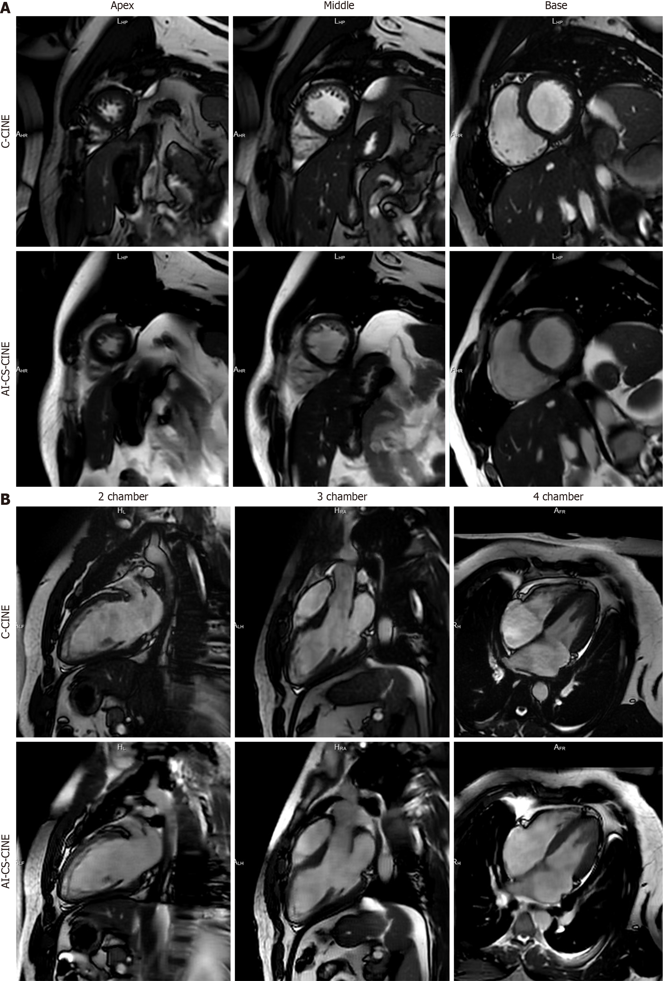
Figure 2 Magnetic resonance imaging illustrating the exceptional alignment between conventional CINE and artificial-intelligence-assisted compressed sensing CINE in a healthy 20-year-old male volunteer.
A: The short-axis stack clearly depicts the apex, middle, and base of the left ventricle in both conventional CINE (C-CINE) (top row) and artificial-intelligence-assisted compressed sensing CINE (AI-CS-CINE) (bottom row); B: Additionally, the 2-, 3-, and 4-chamber views demonstrate excellent agreement between C-CINE and AI-CS-CINE in this volunteer. C-CINE: Conventional CINE; AI-CS-CINE: Artificial-intelligence-assisted compressed sensing CINE.
Figure 3 Exceptional alignment between conventional CINE and artificial-intelligence-assisted compressed sensing CINE is demon
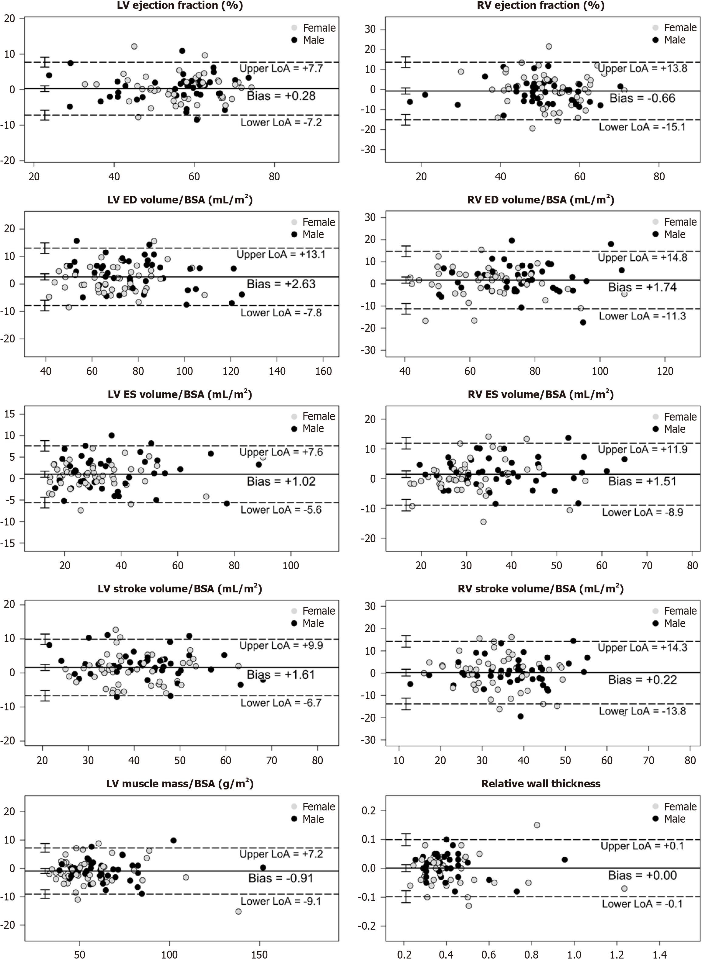
Figure 4 Bland–Altman plots reveal a strong agreement of metrics between conventional CINE and artificial-intelligence-assisted compressed sensing CINE.
The solid line represents the mean difference (bias), while dashed lines denote the upper and lower limits of agreement (LoA1.96SDdiff). Error bars, depicting confidence intervals, are presented for both the average difference and the limits of agreement. Visual inspection of the Bland–Altman plots revealed no obvious relationship between the differences and the magnitude of measurements, nor systematic bias. For each variable, conventional CINE (C-CINE) and artificial-intelligence-assisted compressed sensing CINE (AI-CS-CINE) revealed an acceptable mean difference. Nearly all measurement pairs lie within the limits of agreement, indicating the AI-CS-CINE method is highly comparable to C-CINE. The X-axis represents the mean of C-CINE and AI-CS-CINE values, while the Y-axis shows the difference between C-CINE and AI-CS-CINE. Dashed lines indicate the mean difference and 95% limits of agreement. BSA: Body surface area; LV: Left ventricle; RV: Right ventricle.
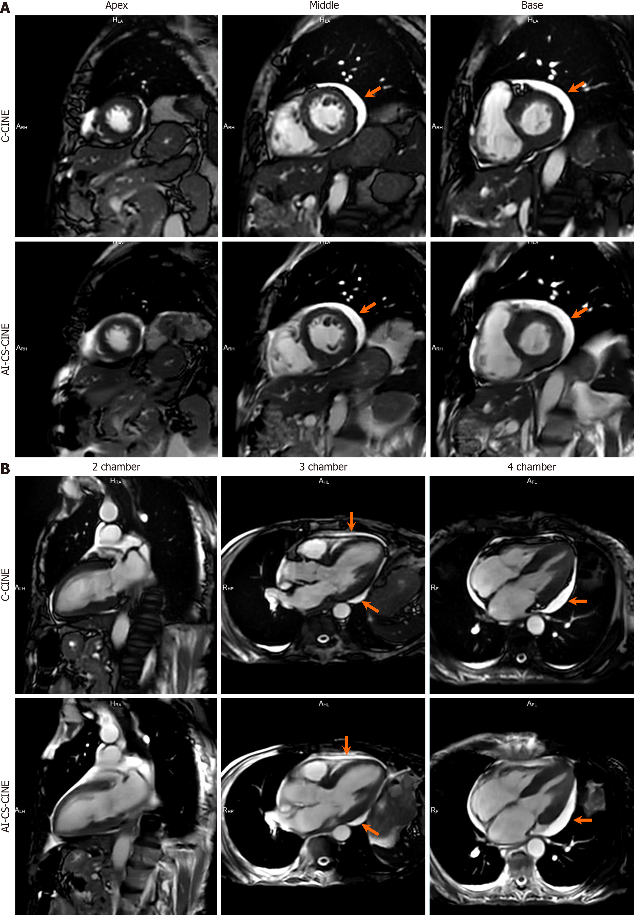
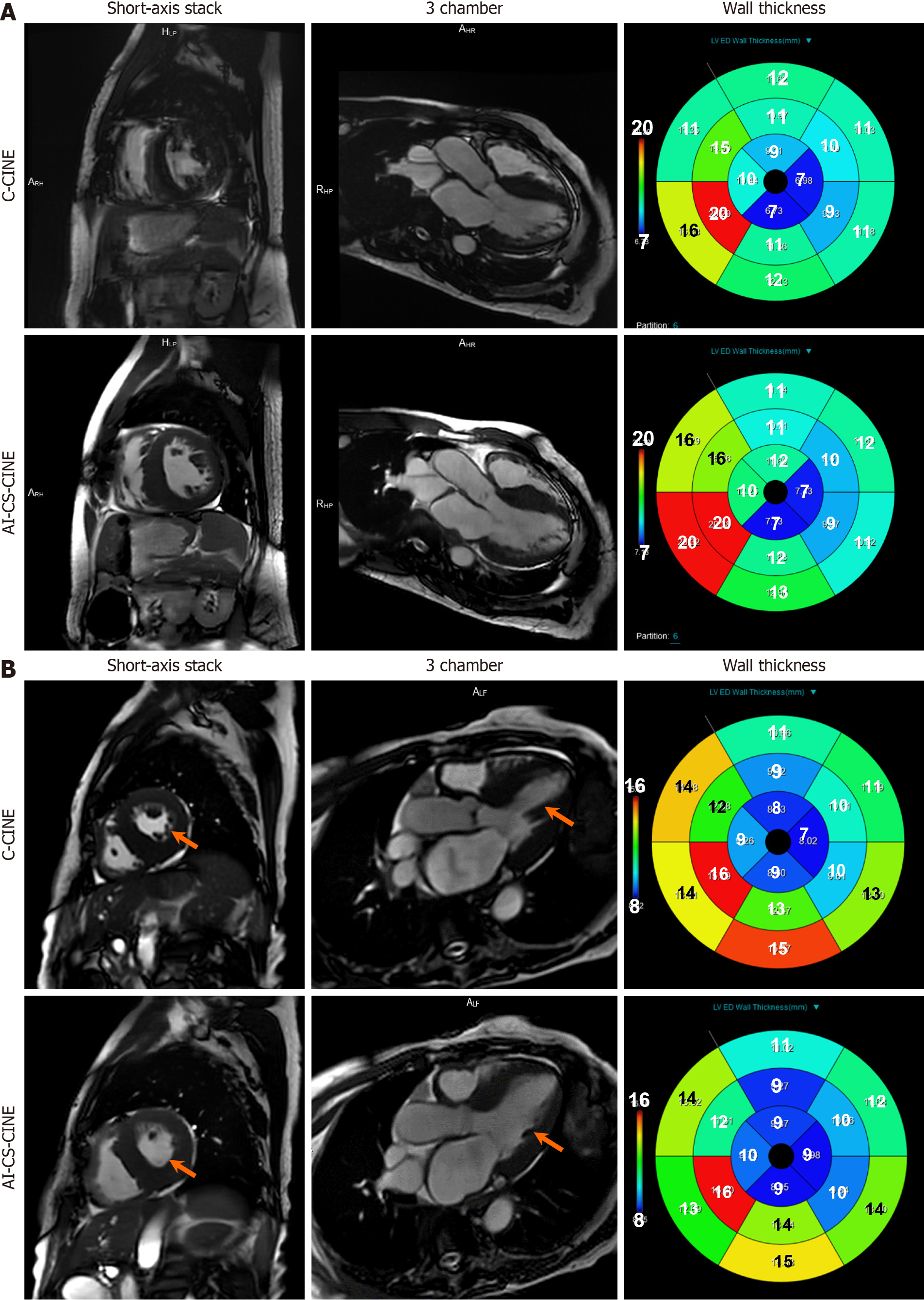
Figure 5 Artificial-intelligence-assisted compressed sensing CINE improved image quality and quantitative reliability in challenging cardiac amyloidosis cases.
A: In a 75-year-old male patient diagnosed with cardiac amyloidosis and small pericardial effusion, poor image quality of the short-axis stack in conventional CINE (C-CINE) was observed due to bad breath holds and poor ECG triggering. These cardiac conditions hindered the patient’s ability to hold his breath for the required 11 seconds during C-CINE acquisition. Consequently, the endomyocardium and perimyocardium were poorly delineated, and trabeculae were not clearly visible (upper row). Such images were considered unsuitable for quantitative image analysis. However, the patient was able to hold his breath during the 2-second artificial-intelligence-assisted compressed sensing CINE (AI-CS-CINE) acquisition, enabling successful image analysis on AI-CS-CINE images only (lower row). Quantitative analysis of wall thickness on C-CINE (upper row) yielded substantially different results compared to AI-CS-CINE (lower row); B: In an 84-year-old female patient diagnosed with cardiac amyloidosis, poor ECG triggering resulted in missing imaging frames during the diastolic phase. Both C-CINE (top row) and AI-CS-CINE (bottom row) exhibited satisfactory image quality for quantitative analysis. However, quantitative metrics did not align well between C-CINE and AI-CS-CINE (right column). Due to inadequate ECG triggering, missing image frames during the diastolic phase were observed in C-CINE, with the end of the full diastolic phase not being displayed (top row). This discrepancy is evident in the 3-chamber view (middle column, arrow). In contrast, the end of the full diastolic phase was successfully displayed in AI-CS-CINE (bottom row, arrow). In this outlying case, AI-CS-CINE outperformed C-CINE, making the metrics derived from AI-CS-CINE more reliable. CINE: Conventional CINE; AI-CS-CINE: Artificial-intelligence-assisted compressed sensing CINE.
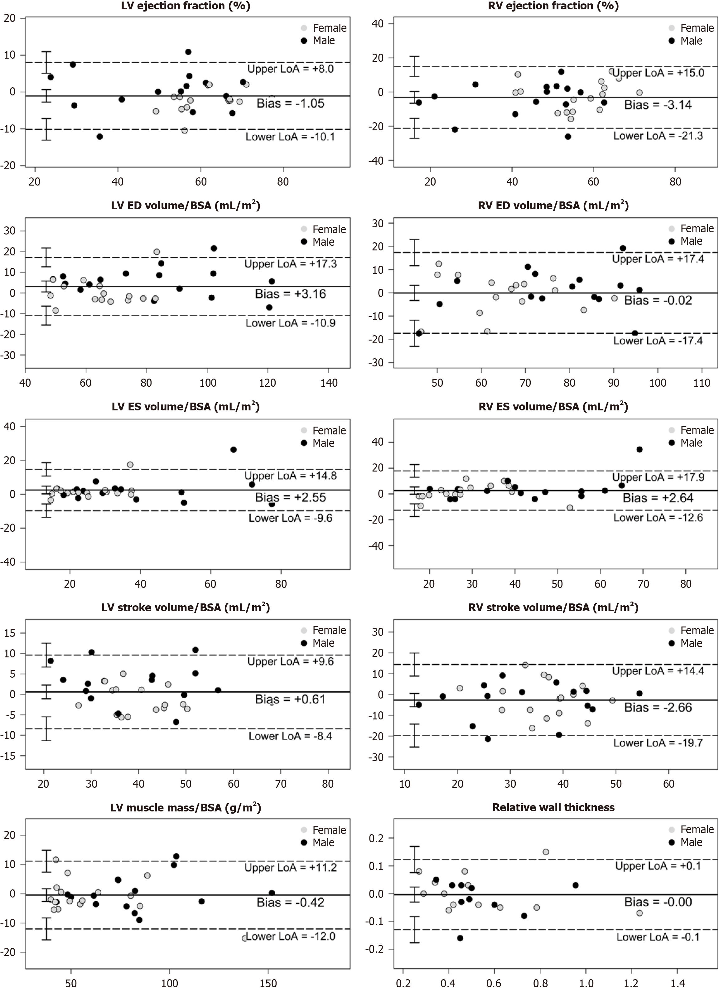
Figure 6 Conventional CINE and artificial-intelligence-assisted compressed sensing CINE still show good correlation in volume and function metrics in left and right ventricle in the subgroup of cardiac amyloidosis (n = 31).
The X-axis represents the mean of conventional CINE (C-CINE) and artificial-intelligence-assisted compressed sensing CINE (AI-CS-CINE) values, while the Y-axis shows the difference between C-CINE and AI-CS-CINE. Dashed lines indicate the mean difference and 95% limits of agreement. BSA: Body surface area; LV: Left ventricle; RV: Right ventricle.
- Citation: Wang H, Schmieder A, Watkins M, Wang P, Mitchell J, Qamer SZ, Lanza G. Artificial intelligence-assisted compressed sensing CINE enhances the workflow of cardiac magnetic resonance in challenging patients. World J Cardiol 2025; 17(7): 108745
- URL: https://www.wjgnet.com/1949-8462/full/v17/i7/108745.htm
- DOI: https://dx.doi.org/10.4330/wjc.v17.i7.108745


