Copyright
©The Author(s) 2025.
World J Cardiol. Jun 26, 2025; 17(6): 106525
Published online Jun 26, 2025. doi: 10.4330/wjc.v17.i6.106525
Published online Jun 26, 2025. doi: 10.4330/wjc.v17.i6.106525
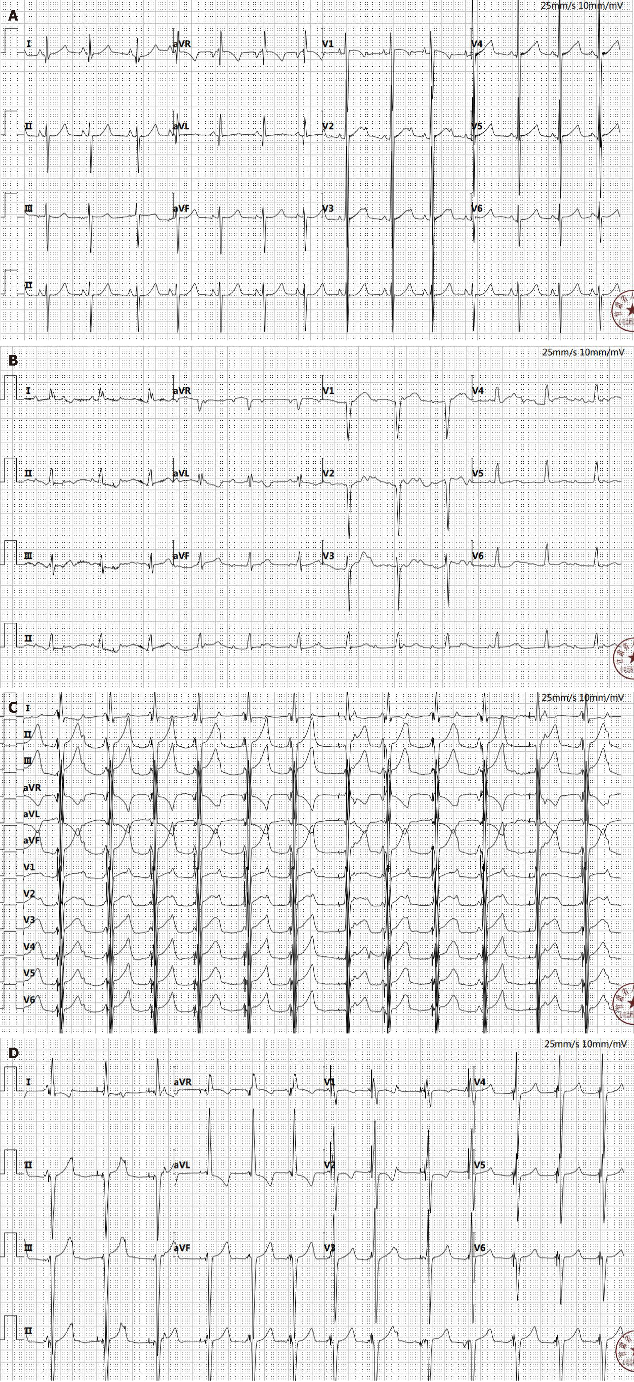
Figure 1 Sequential electrocardiogram changes in the clinical course: from admission to post-discharge follow up.
A: The initial electrocardiogram (ECG) upon admission; B: Postoperative ECG on the first recording after surgical intervention; C: ECG post-permanent pacemaker implantation; D: ECG upon follow-up visit one month after hospital discharge.
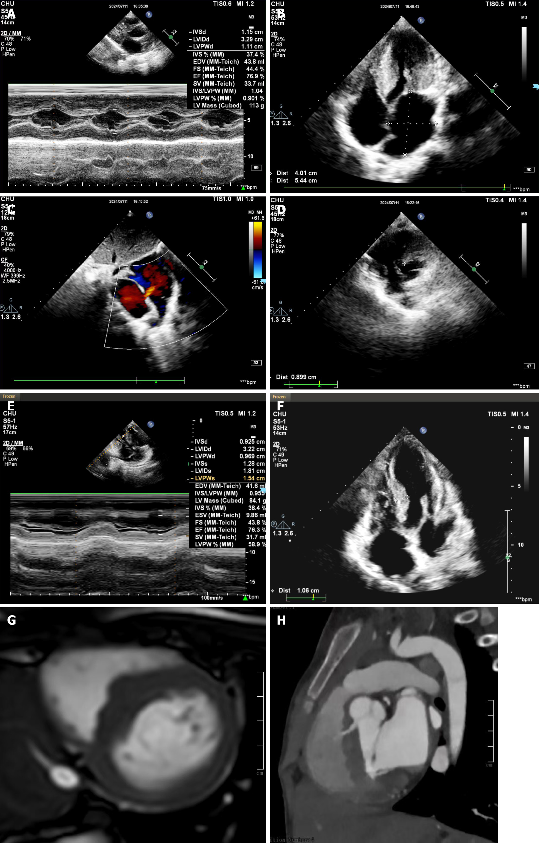
Figure 2 Echocardiographic and cardiac magnetic resonance imaging findings: from pre-operative diagnosis to post-discharge resolution in cardiac pathology.
A: Preoperative echocardiogram indicates significant left ventricular wall thickening; B: Echocardiogram showing dilation left atrium left ventricular outflow tract obstruction; C: Echocardiogram: Atrial septal echo dropout with left-to-right shunt at the atrial level; D: Echocardiogram: Membranous part of the interventricular septum echo dropout measuring 89 mm; E: One month post-discharge, the echocardiogram reveals a septal thickness of 0.925 cm at end-diastole; F: Interventricular septum after alleviation of the obstruction; G: Short-axis cine end-diastolic frame of the heart; H: Cardiac magnetic resonance imaging showing hypertrophied interventricular septum and ventricular septal defect.
- Citation: Ma N, Li ZW, Liu JJ, Liu XG, Zhou X, Wang BW, Li YL, Zhang TC, Xie P. RAF1 mutation expands the cardiac phenotypic spectrum of Noonan syndrome: A case report. World J Cardiol 2025; 17(6): 106525
- URL: https://www.wjgnet.com/1949-8462/full/v17/i6/106525.htm
- DOI: https://dx.doi.org/10.4330/wjc.v17.i6.106525


