Copyright
©The Author(s) 2024.
World J Cardiol. Jun 26, 2024; 16(6): 314-317
Published online Jun 26, 2024. doi: 10.4330/wjc.v16.i6.314
Published online Jun 26, 2024. doi: 10.4330/wjc.v16.i6.314
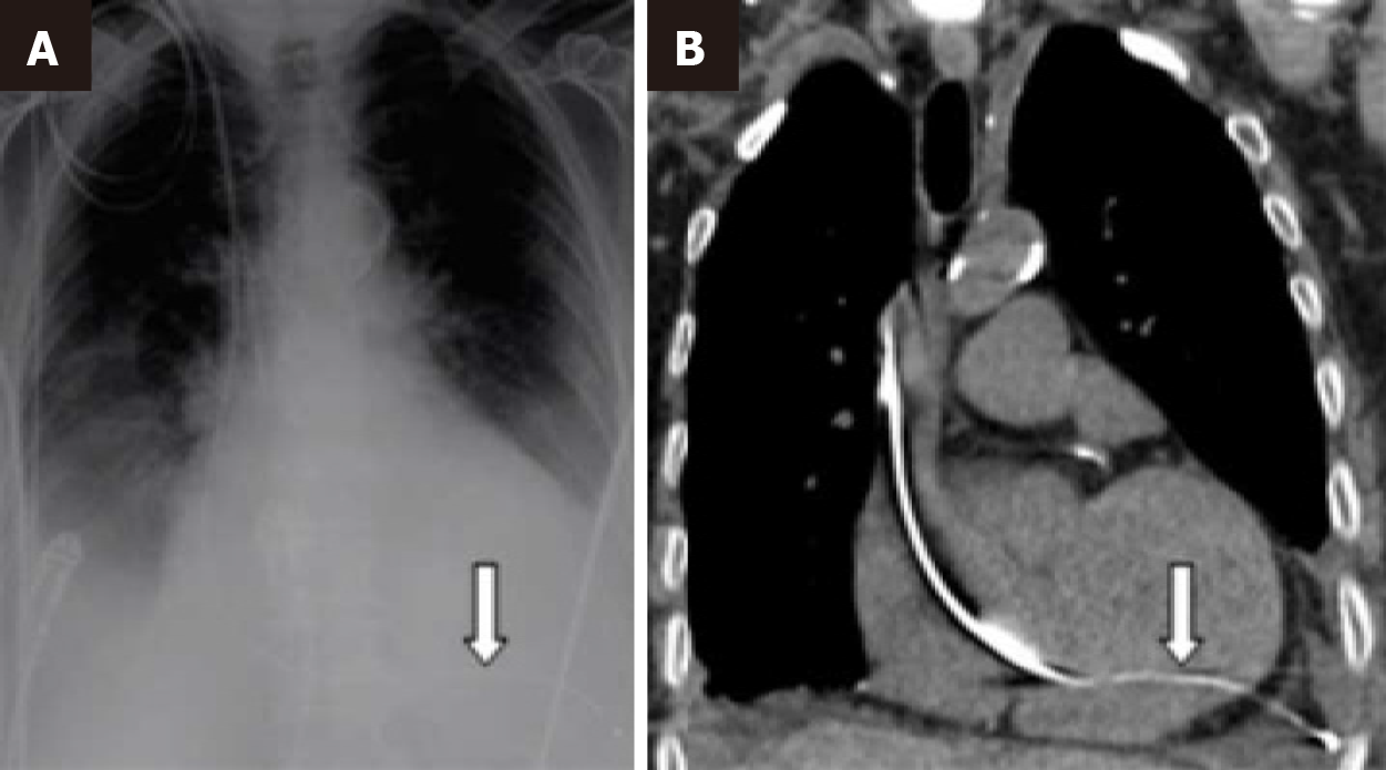
Figure 1 Chest radiography and computed tomography.
A: Chest radiography (arrow); B: Computed tomography (arrow) showing that the pacing wire had traversed from the inter-ventricular septum into the left ventricle, and through the left ventricular myocardium to lie within the left pleura.
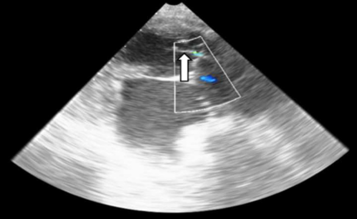
Figure 2 Transoesophageal echocardiogram on admission demonstrated severe Carpentier IIIB mitral regurgitation from chordal entrap
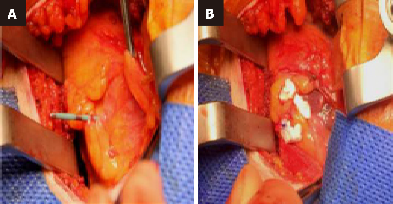
Figure 3 Following left anterior thoracotomy in the 6th intercostal space.
A: A Teflon-pledgeted 3-0 polypropylene purse-string suture was tied around the protruding pacing wire at the left ventricular apex; B: Alongside transvenous lead withdrawal.
- Citation: Acharya M, Kavanagh E, Garg S, Sef D, De Robertis F. Management of a patient with an unusual trajectory of a temporary trans-venous pacing lead. World J Cardiol 2024; 16(6): 314-317
- URL: https://www.wjgnet.com/1949-8462/full/v16/i6/314.htm
- DOI: https://dx.doi.org/10.4330/wjc.v16.i6.314


