Copyright
©The Author(s) 2024.
World J Cardiol. Apr 26, 2024; 16(4): 173-176
Published online Apr 26, 2024. doi: 10.4330/wjc.v16.i4.173
Published online Apr 26, 2024. doi: 10.4330/wjc.v16.i4.173
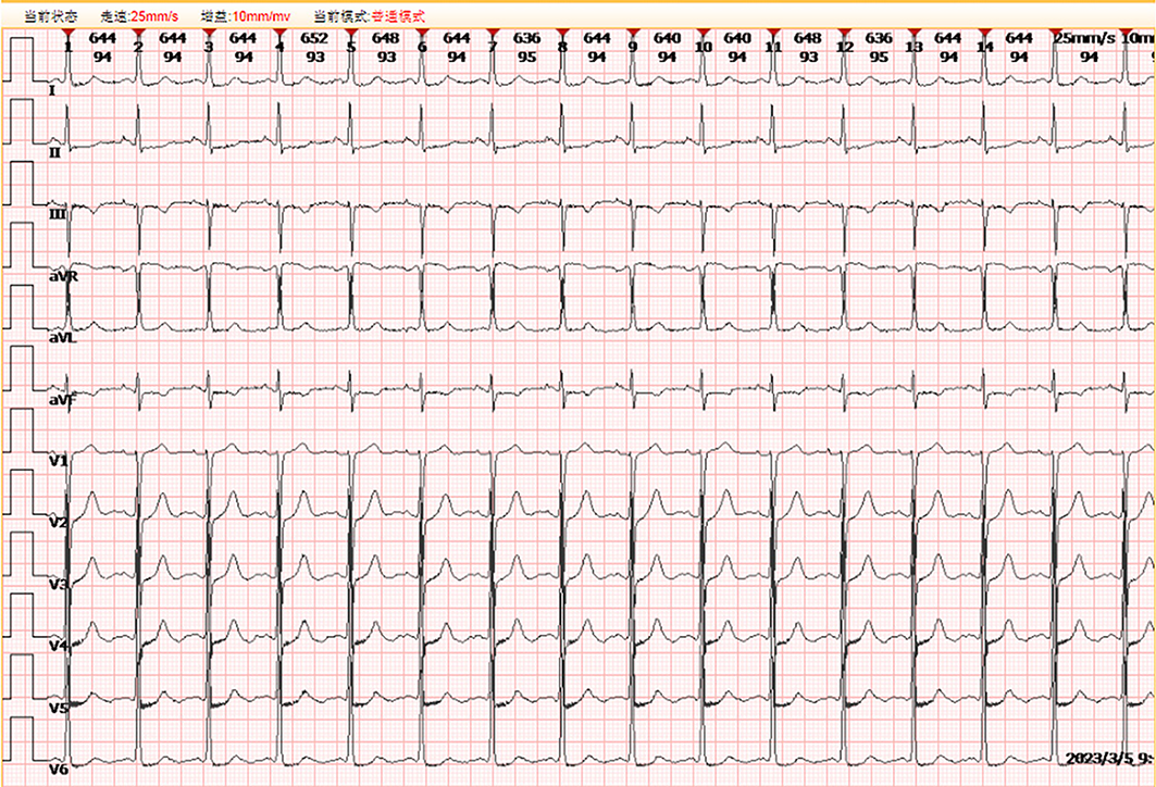
Figure 1 Electrocardiography at initial presentation[1].
Normal sinus rhythm with ST-T wave changes in leads I, II, III, aVF, and V4-V6. Citation: Zhou YP, Wang LL, Qiu YG, Huang SW. R-I subtype single right coronary artery with congenital absence of left coronary system: A case report. World J Cardiol 2023; 15: 649-654. Copyright ©2023 The Authors. Published by Baishideng Publishing Group Inc.
Figure 2 Coronary angiography[1].
A: No coronary artery was found on multiple coronary projections; B and C: A single and large coronary artery originating from the right coronary sinus continued in the coronary sulcus (B), and extended to the anterior base of the heart, where it gave rise to the left anterior descending coronary artery (C); D: No coronary artery was found on a non-selective aortic root injection. Citation: Zhou YP, Wang LL, Qiu YG, Huang SW. R-I subtype single right coronary artery with congenital absence of left coronary system: A case report. World J Cardiol 2023; 15: 649-654. Copyright ©2023 The Authors. Published by Baishideng Publishing Group Inc.
- Citation: Ito S. Challenging situation of coronary artery anomaly associated with ischemia and/or risk of sudden death. World J Cardiol 2024; 16(4): 173-176
- URL: https://www.wjgnet.com/1949-8462/full/v16/i4/173.htm
- DOI: https://dx.doi.org/10.4330/wjc.v16.i4.173


