Copyright
©The Author(s) 2024.
World J Cardiol. Feb 26, 2024; 16(2): 73-79
Published online Feb 26, 2024. doi: 10.4330/wjc.v16.i2.73
Published online Feb 26, 2024. doi: 10.4330/wjc.v16.i2.73
Figure 1 Estimation of right atrial pressure by inferior vena cava ultrasound in spontaneously breathing patients, based on current guidelines[8].
IVC: Inferior vena cava; RA: Right atrium; HV: Hepatic vein; RAP: Rright atrial pressure.
Figure 2 Internal jugular vein collapse point compared to a wine bottle and paint brush.
Figures adapted from NephroPOCUS.com with permission.
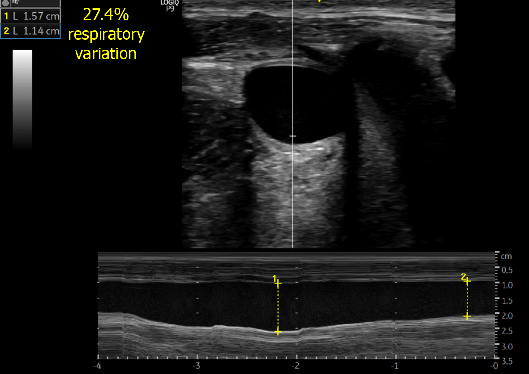
Figure 3 Anteroposterior diameter of the internal jugular vein.
M-mode tracing depicts respiratory variation in the diameter.
Figure 4 Increase in the size of internal jugular vein with Valsalva maneuver by several folds in a spontaneously breathing person with normal right atrial pressure.
Figure 5 Minimal increase in the size of internal jugular vein with Valsalva maneuver in a spontaneously breathing heart failure patient with elevated right atrial pressure.
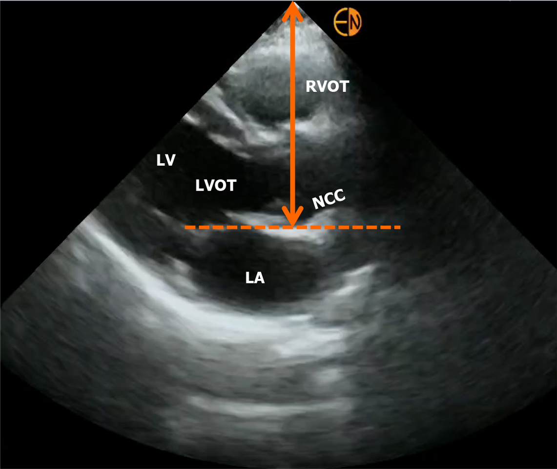
Figure 6 Measurement of right atrial depth using parasternal long axis view on focused cardiac ultrasound.
RVOT: Right ventricular outflow tract; LV: Left ventricle; LVOT: LV outflow tract; LA: Left atrium; NCC: Non-coronary cusp of aortic valve.
Figure 7 Diagnostic algorithm in a case of cirrhosis and suspected hemodynamic acute kidney injury.
Incorrect angle of insonation is a frequent source of error when assessing LVOT VTI (surrogate for stroke volume) and other Doppler measurements listed. Adapted from Ref. 33 with kind permission of the publisher (corresponding author’s prior open access publication). Blue boxes: Right heart; Red boxes: Left heart-related sonographic parameters; Green outlines: Volume tolerance phenotype; Orange outlines: Volume intolerance. POCUS: Point-of-care ultrasonography; VTI: Velocity time integral; E/e′: Ratio of the early diastolic waves of the mitral inflow Doppler and mitral annular tissue Doppler; LA: Left atrium; RV: Right ventricle; RVSP: Right ventricular systolic pressure; TAPSE: Tricuspid annular plane systolic excursion; S′: Tricuspid annular systolic velocity; SVC: Superior vena cava; ARDS: Acute respiratory distress syndrome; HV: Hepatic vein; PV: Portal vein.
- Citation: Chayapinun V, Koratala A, Assavapokee T. Seeing beneath the surface: Harnessing point-of-care ultrasound for internal jugular vein evaluation. World J Cardiol 2024; 16(2): 73-79
- URL: https://www.wjgnet.com/1949-8462/full/v16/i2/73.htm
- DOI: https://dx.doi.org/10.4330/wjc.v16.i2.73


