Copyright
©The Author(s) 2024.
World J Cardiol. Dec 26, 2024; 16(12): 760-767
Published online Dec 26, 2024. doi: 10.4330/wjc.v16.i12.760
Published online Dec 26, 2024. doi: 10.4330/wjc.v16.i12.760
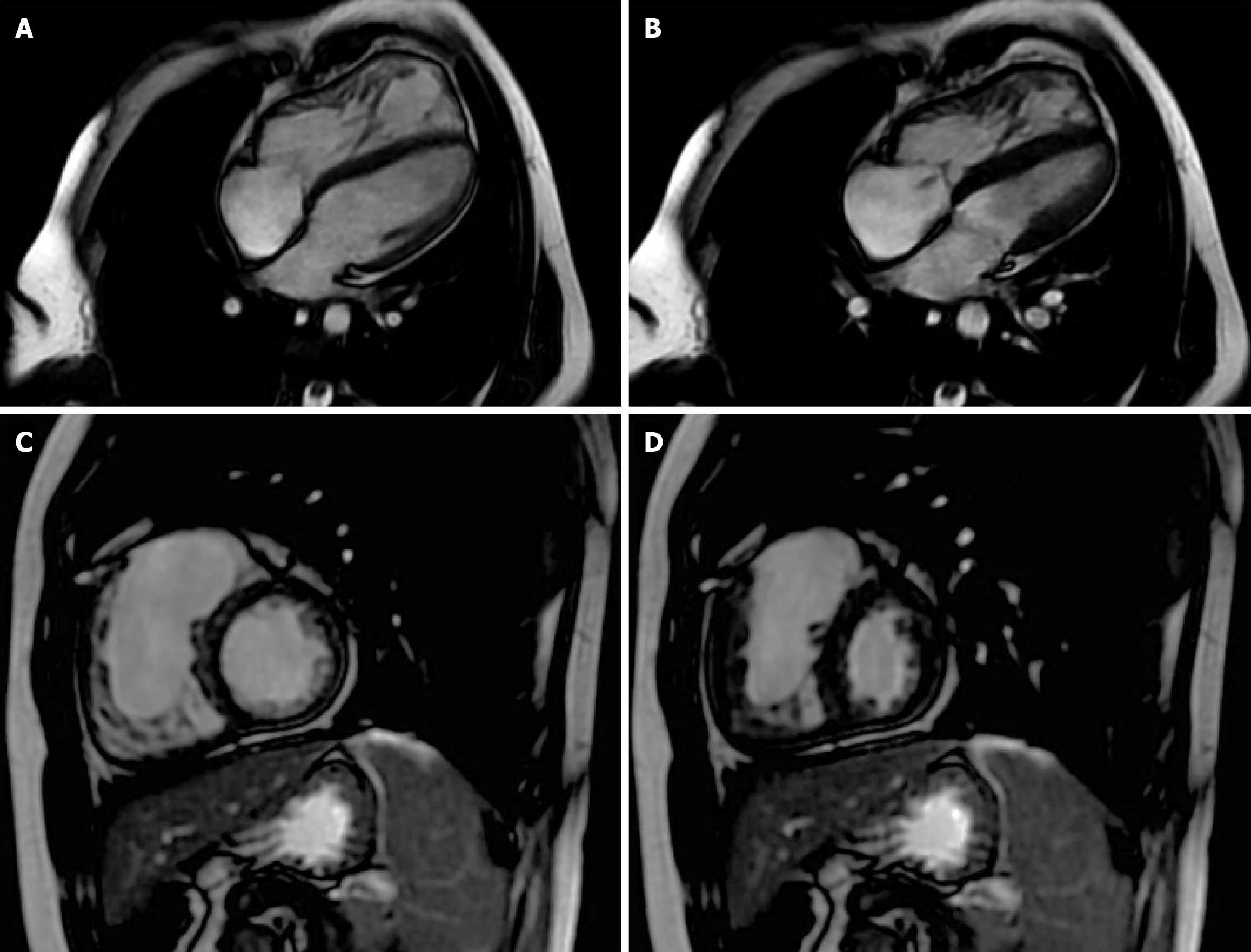
Figure 1 Cardiac magnetic resonance imaging cine sequences (June 2022).
A: Four chamber view: Enlargement of the right ventricle and increased right ventricle trabeculation; B: Systolic image showing tricuspid valve regurgitation; C: Short axis in diastole; D: Short axis in systole. No saccular lesion of the right ventricle was observed in this study.
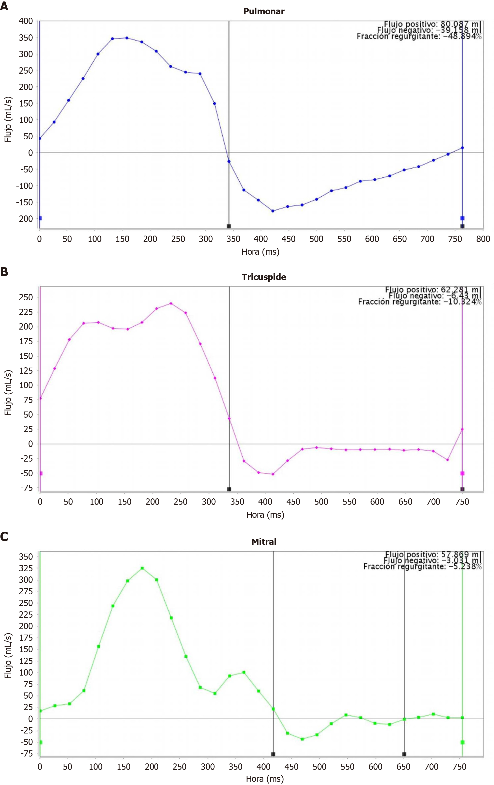
Figure 2 Cardiac magnetic resonance (June 2022).
A: Flow-versus-time graph shows valvular pulmonary insufficiency a regurgitant volume of 39.1 mL per beat and regurgitant fraction of 48.8%; B: Valvular tricuspid insufficiency; a regurgitant volume of 6.4 mL per beat and regurgitant fraction of 10.3%; C: Cardiac magnetic resonance (June 2024). Flow-versus-time graph shows paravalvular leak a regurgitant volume of 4.8 mL per beat and regurgitant fraction 14.8%.
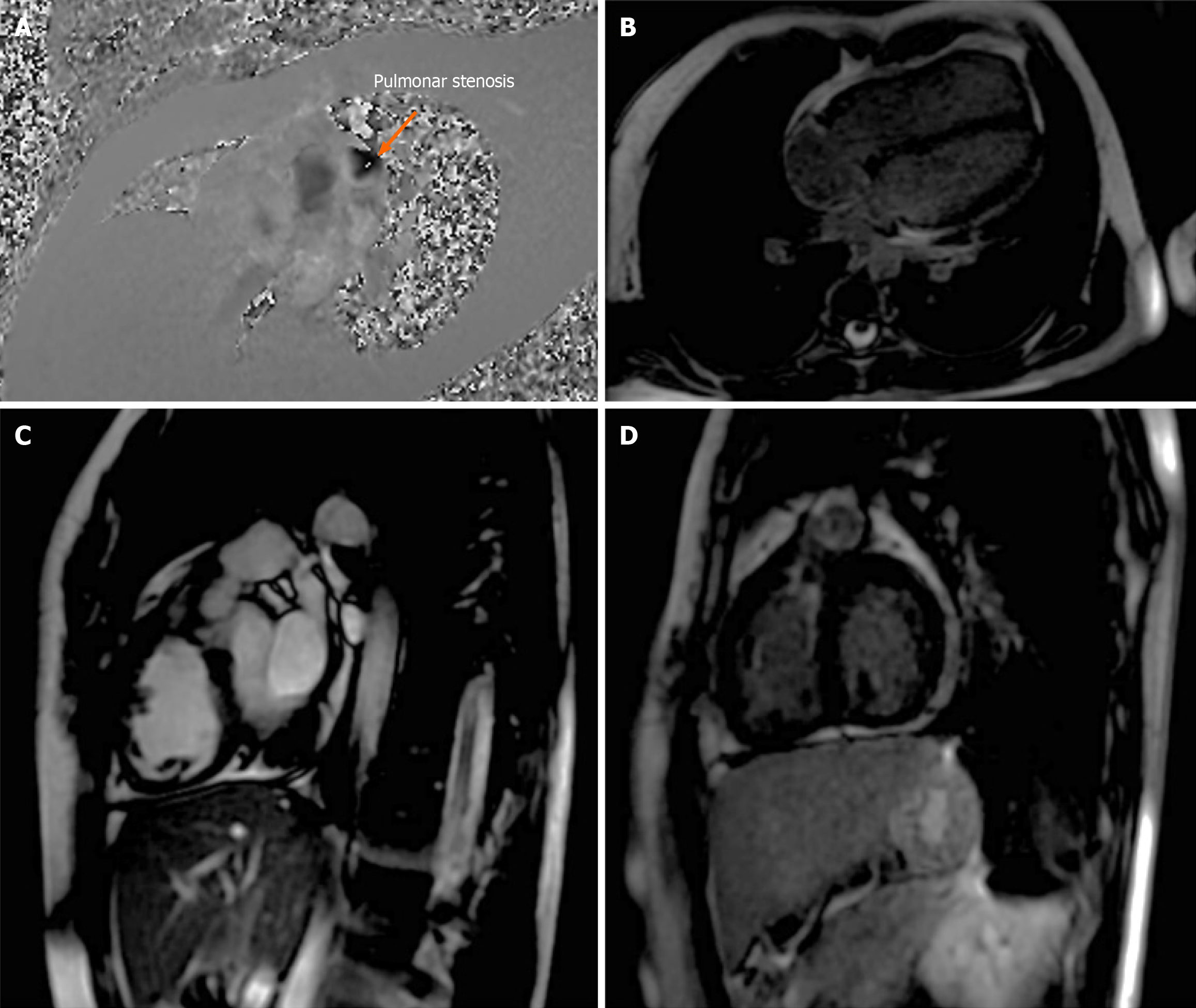
Figure 3 Cardiac magnetic resonance cine sequences (June 2022).
A: Phase-contrast imaging; Right ventricular outflow tract stenosis and increased velocity; B and D: Late gadolinium enhancement sequence; Right atrial enhancement and right ventricular outflow tract; C: Cine sequence; Right ventricular outflow tract obstruction.
Figure 4 Cardiac magnetic resonance (February 2024) diverticulum of the inferolateral wall of the right ventricle after pulmonary valve prosthesis.
A: Four chamber view: Diastole; B: Four chamber view: Systole; C: Four chamber saccular image in the inferolateral wall of the right ventricle, which showed synchronous movement and contraction like the rest of the myocardium.
Figure 5 Cardiac magnetic resonance control right ventricular outflow tract cine sequences, T2 weight.
A: Phase-contrast imaging; Paravalvular leak; B and C: After pulmonary valve placement (February 2024).
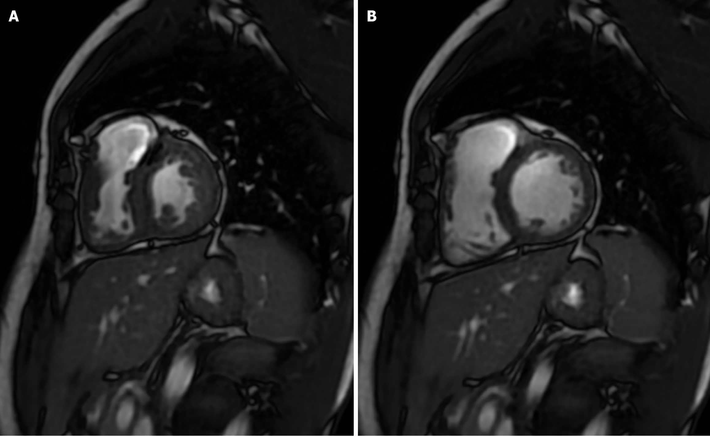
Figure 6 Cardiac magnetic resonance cine sequences short axis (February 2024) normal right ventricle.
A: Systole; B: Diastole.
- Citation: Martinez Juarez D, Gomez Monterrosas O, Tlecuitl Mendoza A, Zamora Rosales F, Álvarez Calderón R, Cepeda Ortiz DA, Espinosa Solis EE. Right ventricular diverticulum following a pulmonary valve placement for correction of tetralogy of Fallot: A case report. World J Cardiol 2024; 16(12): 760-767
- URL: https://www.wjgnet.com/1949-8462/full/v16/i12/760.htm
- DOI: https://dx.doi.org/10.4330/wjc.v16.i12.760


