Copyright
©The Author(s) 2024.
World J Cardiol. Oct 26, 2024; 16(10): 595-603
Published online Oct 26, 2024. doi: 10.4330/wjc.v16.i10.595
Published online Oct 26, 2024. doi: 10.4330/wjc.v16.i10.595
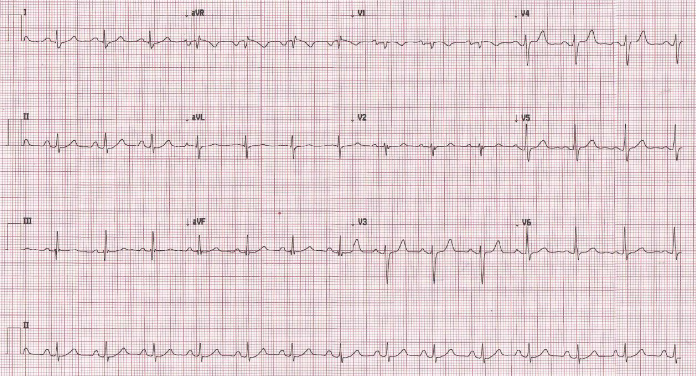
Figure 1 Pre-operative electrocardiogram.
It shows normal sinus rhythm with normal P wave axis and interatrial conduction delay.
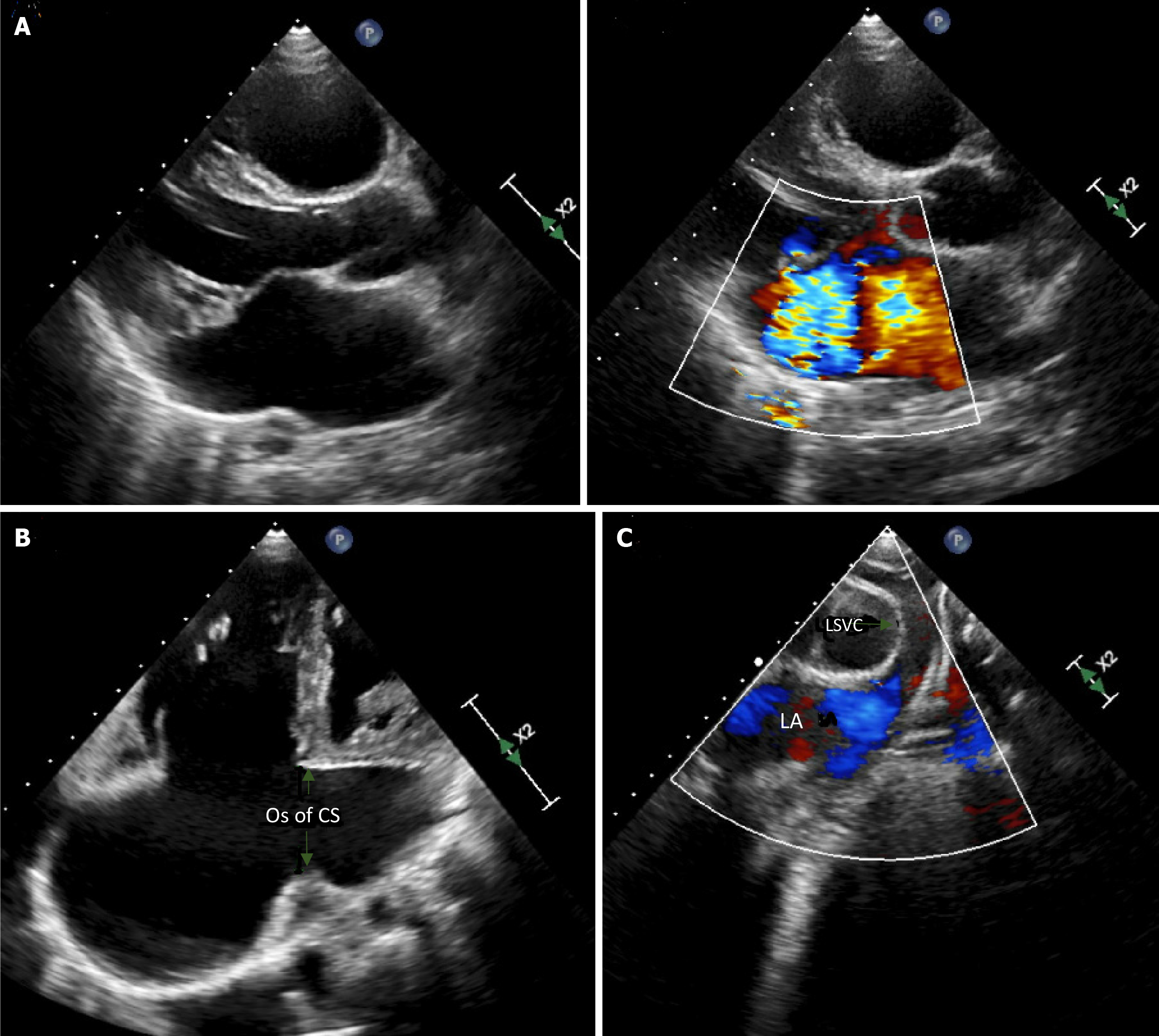
Figure 2 Echocardiographic imaging of the mitral valve.
A: A parasternal long axis view of the mitral valve showing myxomatous prolapsing leaflets and severe mitral regurgitation; B: A modified 4 chamber view, with posterior angulation showing the dilated ostium of the coronary sinus; C: Modified supra coronal cut from the supra-sternal window showing a left superior vena cava draining into the left atrium. OS: Ostium; CS: Coronary sinus; LSVC: Left superior vena cava; LA: Left atrium.
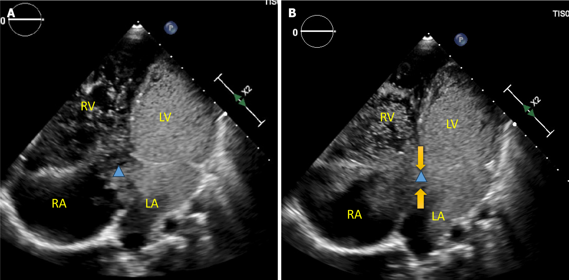
Figure 3 Agitated saline contrast injection in the left antecubital vein.
A: Views taken from the 4-chamber window. Note the contrast filling the left atrium and the left ventricle before crossing the dilated ostium of the unroofed coronary sinus (triangle); B: Arrows pointing to the edges of the coronary sinus ostium. RA: Right atrium; LA: Left atrium; RV: Right ventricle; LV: Left ventricle.
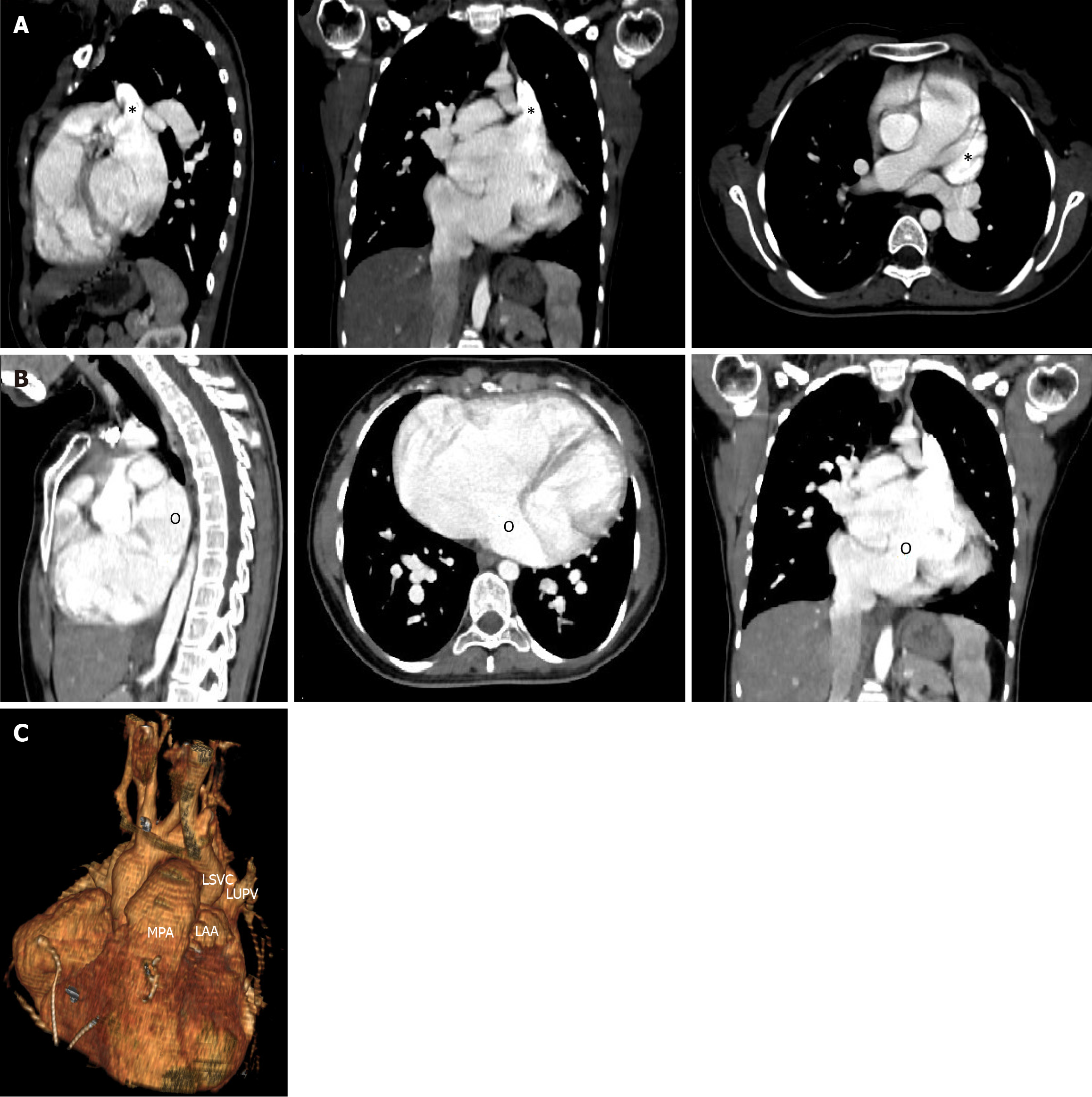
Figure 4 Preoperative computed tomography angiogram.
A: The insertion site of the left superior vena cava (LSVC) is shown by an asterisk in the axial, sagittal and coronal views; B: The ostium of the coronary sinus indicated by the letter (o) is shown in the same three views; C: A three-dimensional reconstruction of the LSCV as it enters the left atrium. LSVC: Left superior vena cava; MPA: Main pulmonary artery; LAA: Left atrial appendage; LUPV: Left upper pulmonary vein.
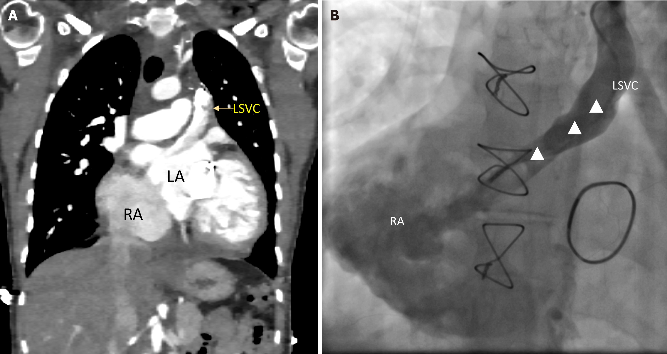
Figure 5 Post-operative imaging.
A: Post-operative computed tomography angiogram; B: Antero-posterior venogram of the left superior vena cava (LSVC). Both images show the course of the intra-atrial tunnel (triangle) connecting the LSVC to the right atrium. RA: Right atrium. LA: Left atrium. LSVC: Left superior vena cava.
- Citation: Bitar F, Bulbul Z, Jassar Y, Zareef R, Abboud J, Arabi M, Bitar FF. Unroofed coronary sinus, left-sided superior vena cava and mitral insufficiency: A case report and review of the literature. World J Cardiol 2024; 16(10): 595-603
- URL: https://www.wjgnet.com/1949-8462/full/v16/i10/595.htm
- DOI: https://dx.doi.org/10.4330/wjc.v16.i10.595


