Copyright
©The Author(s) 2023.
World J Cardiol. May 26, 2023; 15(5): 244-252
Published online May 26, 2023. doi: 10.4330/wjc.v15.i5.244
Published online May 26, 2023. doi: 10.4330/wjc.v15.i5.244
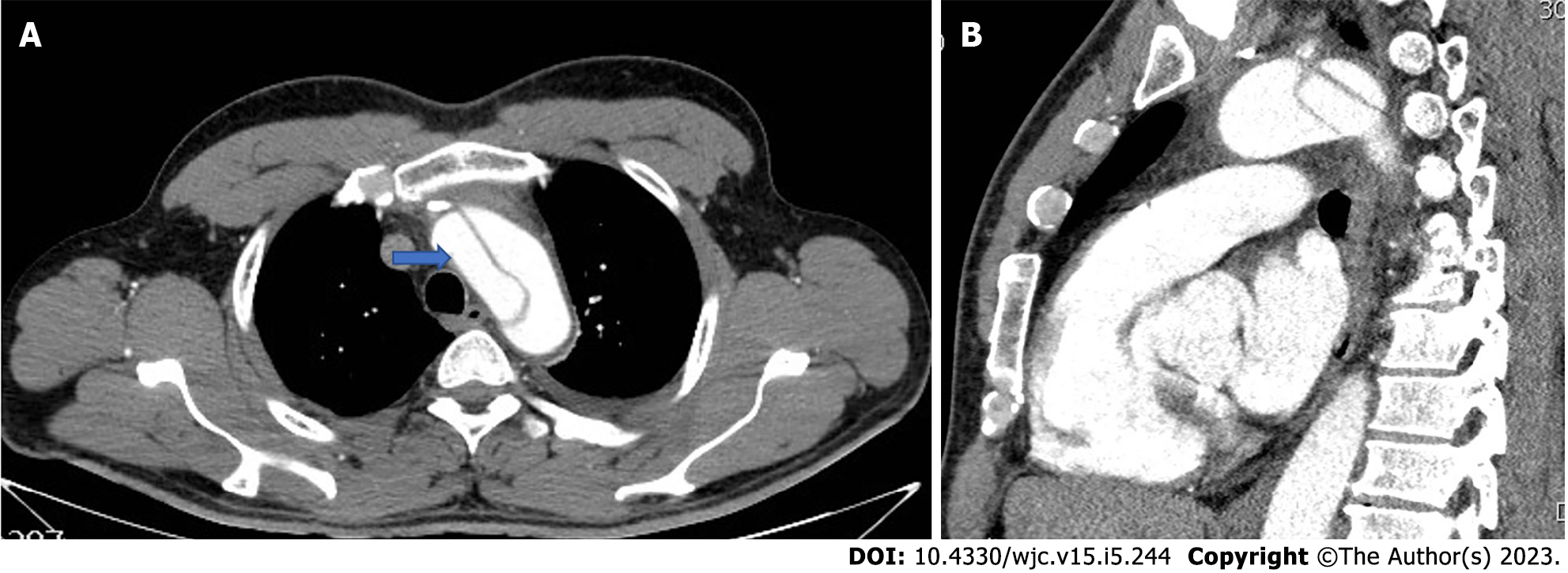
Figure 1 Pre-operative computed tomography images of a non-A non-B aortic dissection case contained in the aortic arch, which was surgically managed in our department.
A: Axial view, blue arrow shows the true lumen; B: Sagittal view.
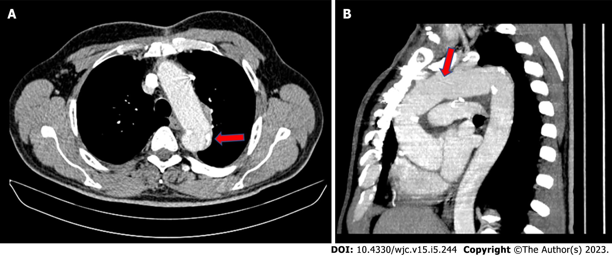
Figure 2 The aortic arch was replaced with a graft (prefabricated aortic branched graft), which contained three pre-attached branches for all three great vessels.
A: Axial view, red arrow demarcates distal anastomotic suture line; B: Sagittal view; red arrow the proximal suture line with the ascending aorta.
- Citation: Christodoulou KC, Karangelis D, Efenti GM, Sdrevanos P, Browning JR, Konstantinou F, Georgakarakos E, Mitropoulos FA, Mikroulis D. Current knowledge and contemporary management of non-A non-B aortic dissections. World J Cardiol 2023; 15(5): 244-252
- URL: https://www.wjgnet.com/1949-8462/full/v15/i5/244.htm
- DOI: https://dx.doi.org/10.4330/wjc.v15.i5.244


