Copyright
©The Author(s) 2023.
World J Cardiol. Oct 26, 2023; 15(10): 542-552
Published online Oct 26, 2023. doi: 10.4330/wjc.v15.i10.542
Published online Oct 26, 2023. doi: 10.4330/wjc.v15.i10.542
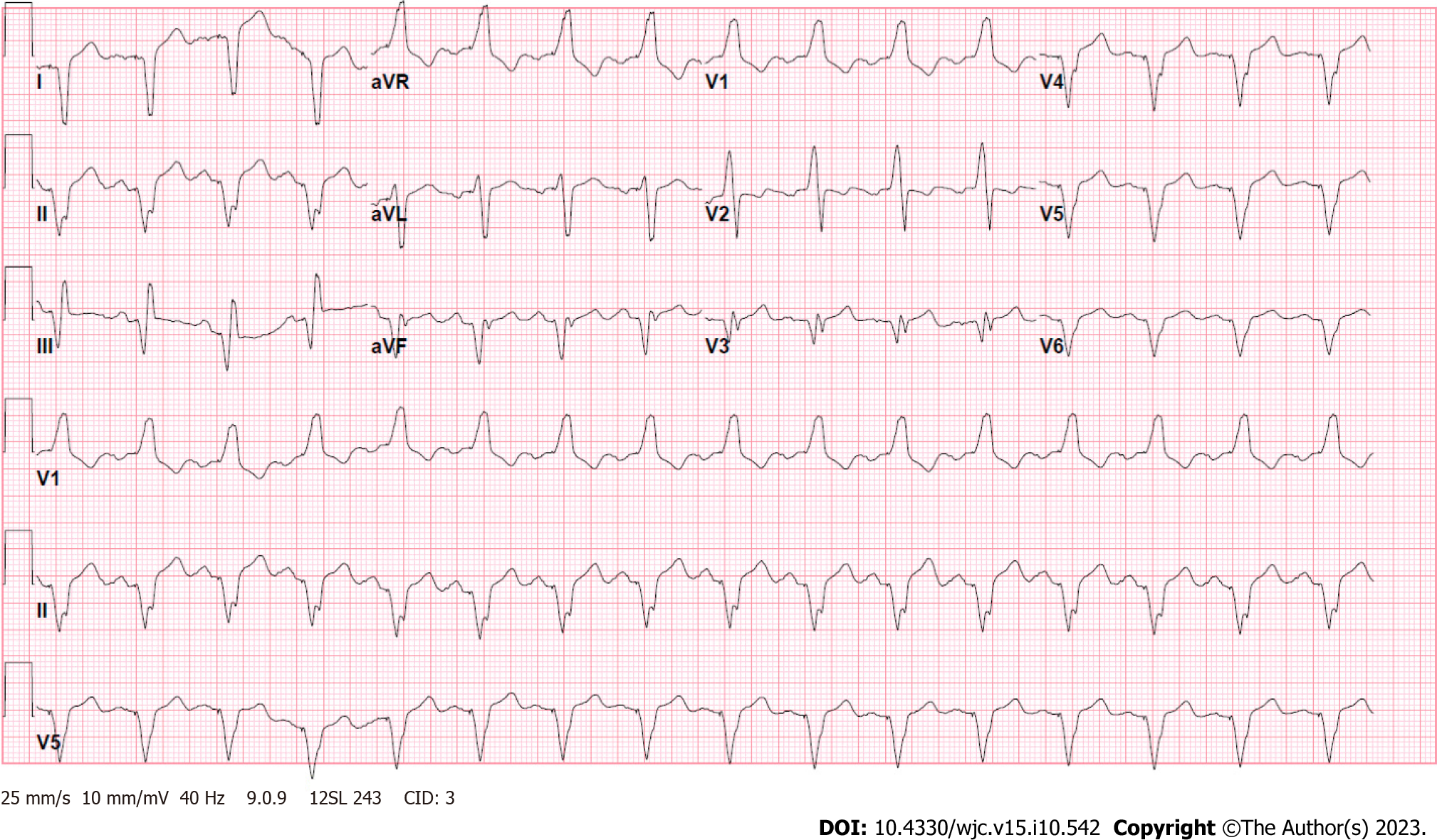
Figure 1 Electrocardiogram showing an atrial-sensed ventricularly-paced rhythm without acute abnormalities.
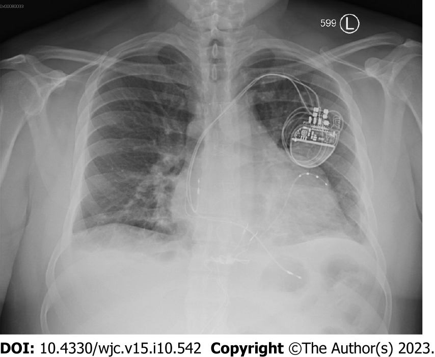
Figure 2 Chest X-ray showing cardiomegaly with small bilateral pleural effusions with unremarkable pulmonary vasculature.
Biventricular pacemaker is present at the right chest.
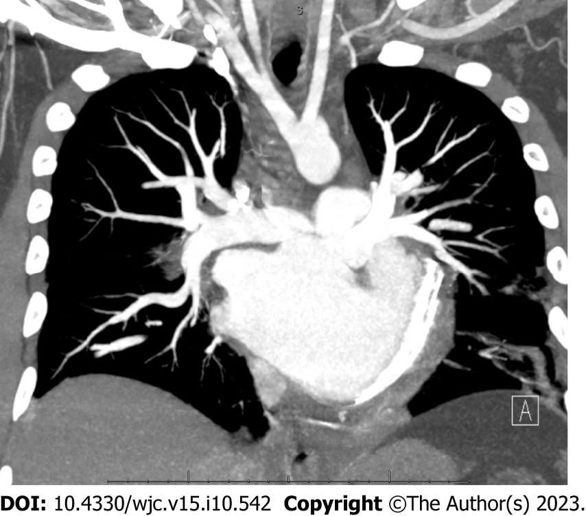
Figure 3 Chest pulmonary angiography demonstrating a levo-transposition of the great arteries cardiac anatomy.
Examination was negative for pulmonary embolism, pericardial effusion, or pneumothorax.
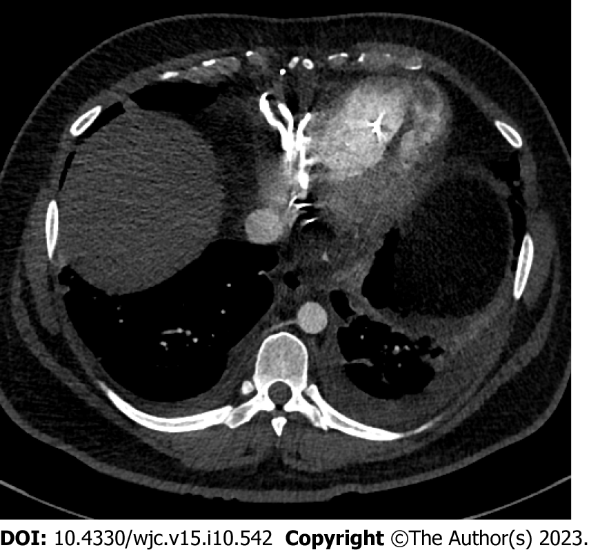
Figure 4 Computed tomography of the chest detailing the patient’s cardiac anatomy of levo-transposition of the great arteries.
Venous circulation consists of the right atrium, left ventricle, and pulmonary artery. Systemic circulation consists of the pulmonary vein, left atrium, right ventricle, and aorta. Patient has a left-sided aortic arch with typical configuration.
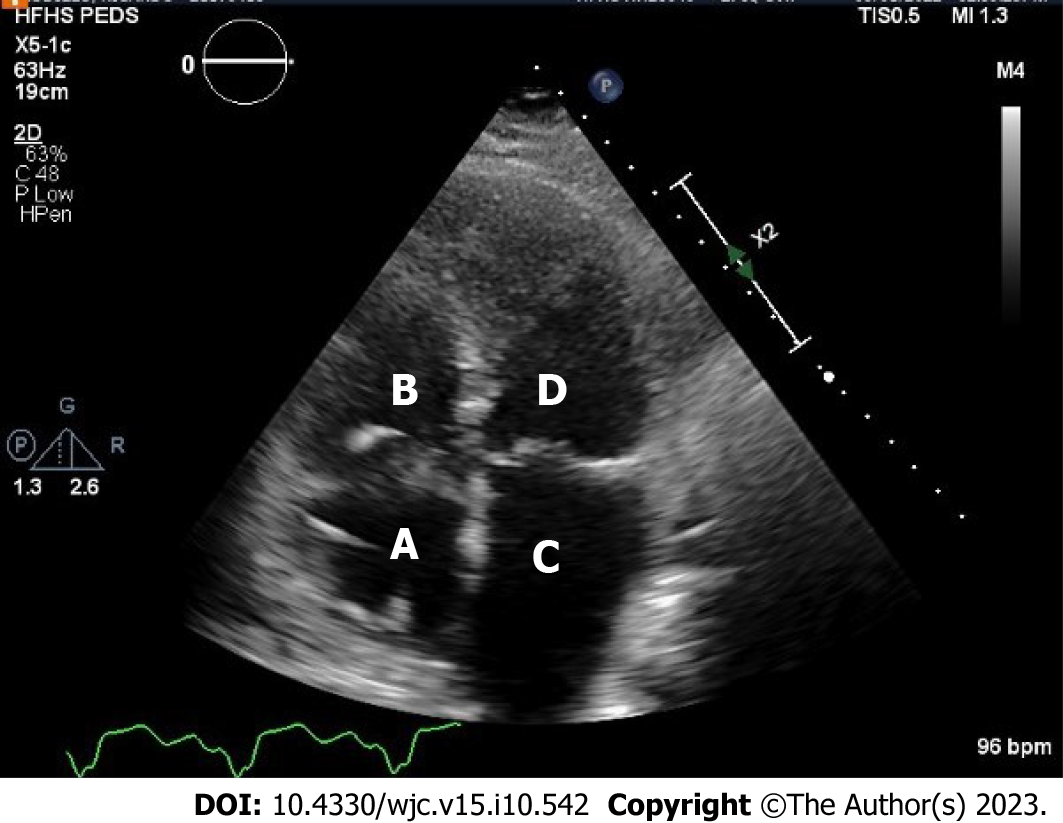
Figure 5 Transthoracic echocardiography demonstrating the patient’s classic levo-transposition of the great arteries anatomy.
A: Right atrium; B: Venous left ventricle; C: Left atrium; D: Systemic right ventricle. Visualized is an apically displaced systemic tricuspid valve opening into systemic right ventricle.
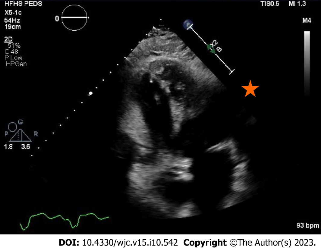
Figure 6 Transthoracic echocardiography in apical four chamber view showing an systemic right ventricle apical thrombus.
This view highlights the importance of visualizing the true apex of the systemic right ventricle as the thrombus is not seen in Figure 5 where the apex is foreshortened.
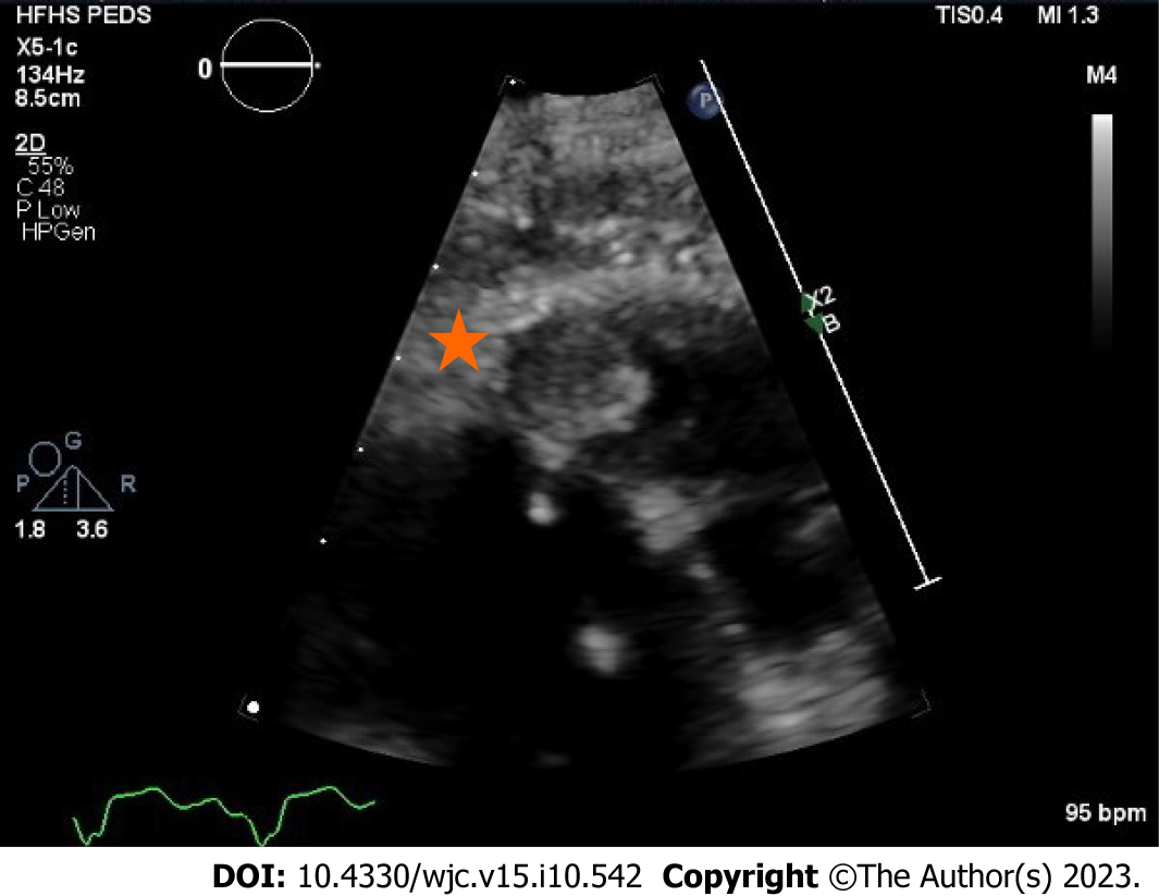
Figure 7 Transthoracic echocardiography highlighting the systemic right ventricle apical thrombus.
A 1.3 cm × 1.3 cm well circumscribed mass with an echolucent center.
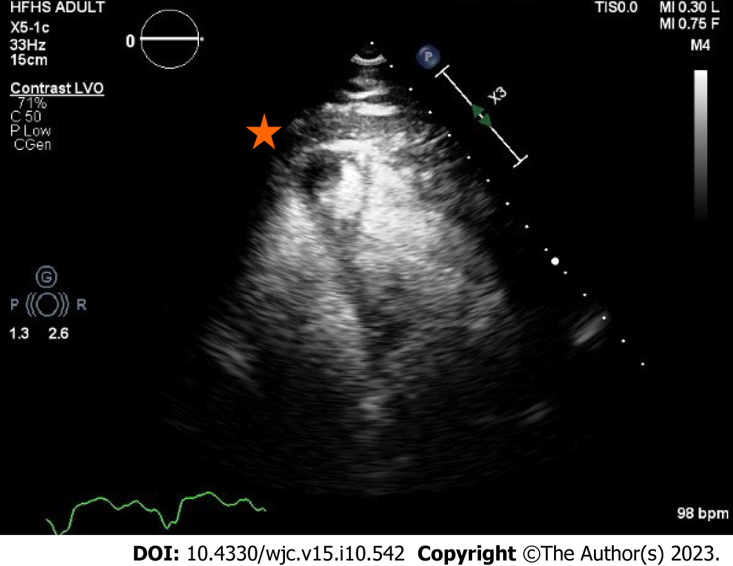
Figure 8 Transthoracic echocardiography with intravenous contrast demarcating the apical thrombus in the systemic right ventricle.
- Citation: Almajed MR, Almajed A, Khan N, Obri MS, Ananthasubramaniam K. Systemic right ventricle complications in levo-transposition of the great arteries: A case report and review of literature. World J Cardiol 2023; 15(10): 542-552
- URL: https://www.wjgnet.com/1949-8462/full/v15/i10/542.htm
- DOI: https://dx.doi.org/10.4330/wjc.v15.i10.542


