Copyright
©The Author(s) 2022.
World J Cardiol. May 26, 2022; 14(5): 297-306
Published online May 26, 2022. doi: 10.4330/wjc.v14.i5.297
Published online May 26, 2022. doi: 10.4330/wjc.v14.i5.297
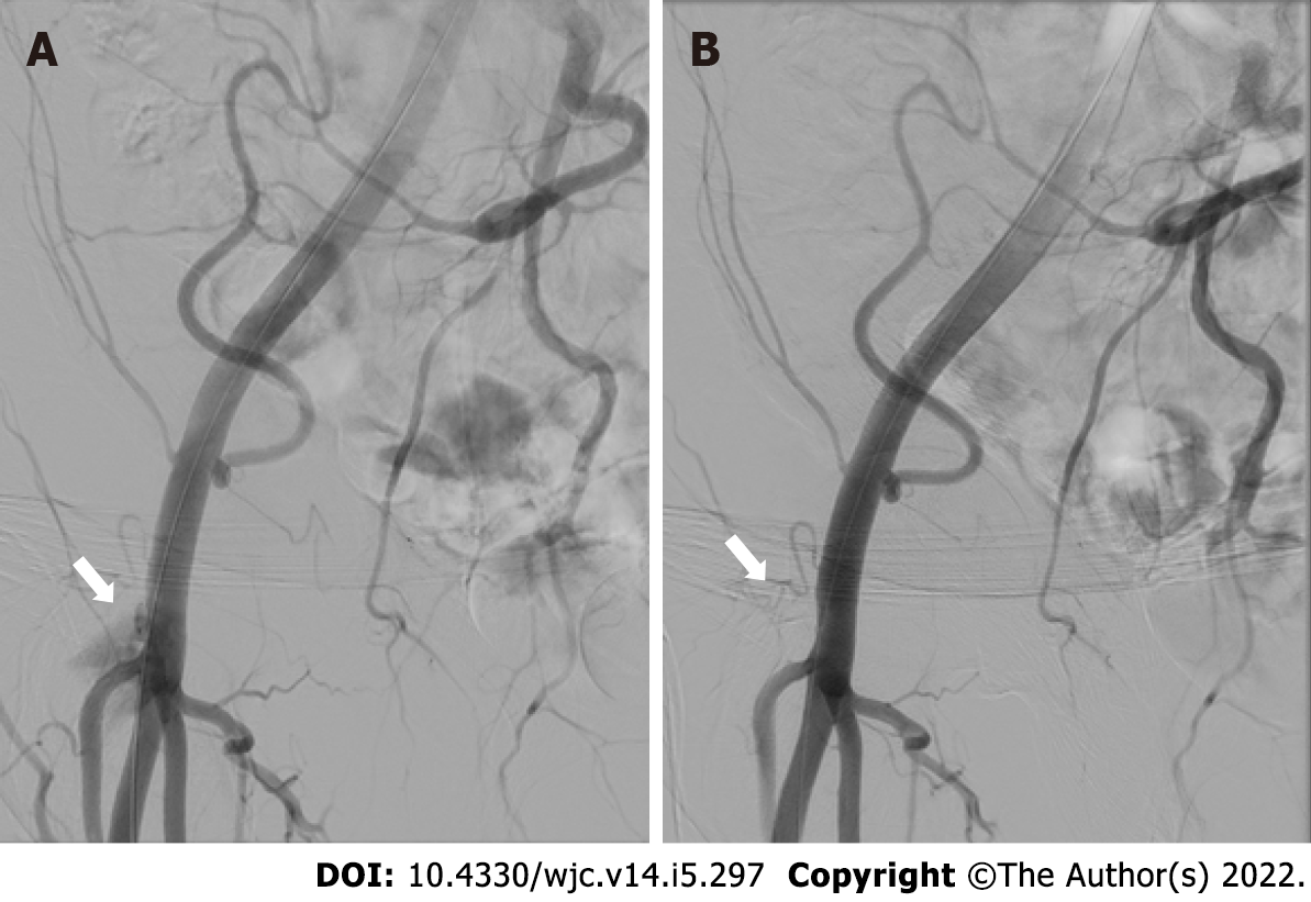
Figure 1 Angiography before and after pledget-assisted hemostasis.
A: Residual bleeding at the transcatheter-aortic-valve-replacement access site (white arrow) after double ProGlide preclosure; B: Absence of residual bleeding (white arrow) after pledget assisted hemostasis.
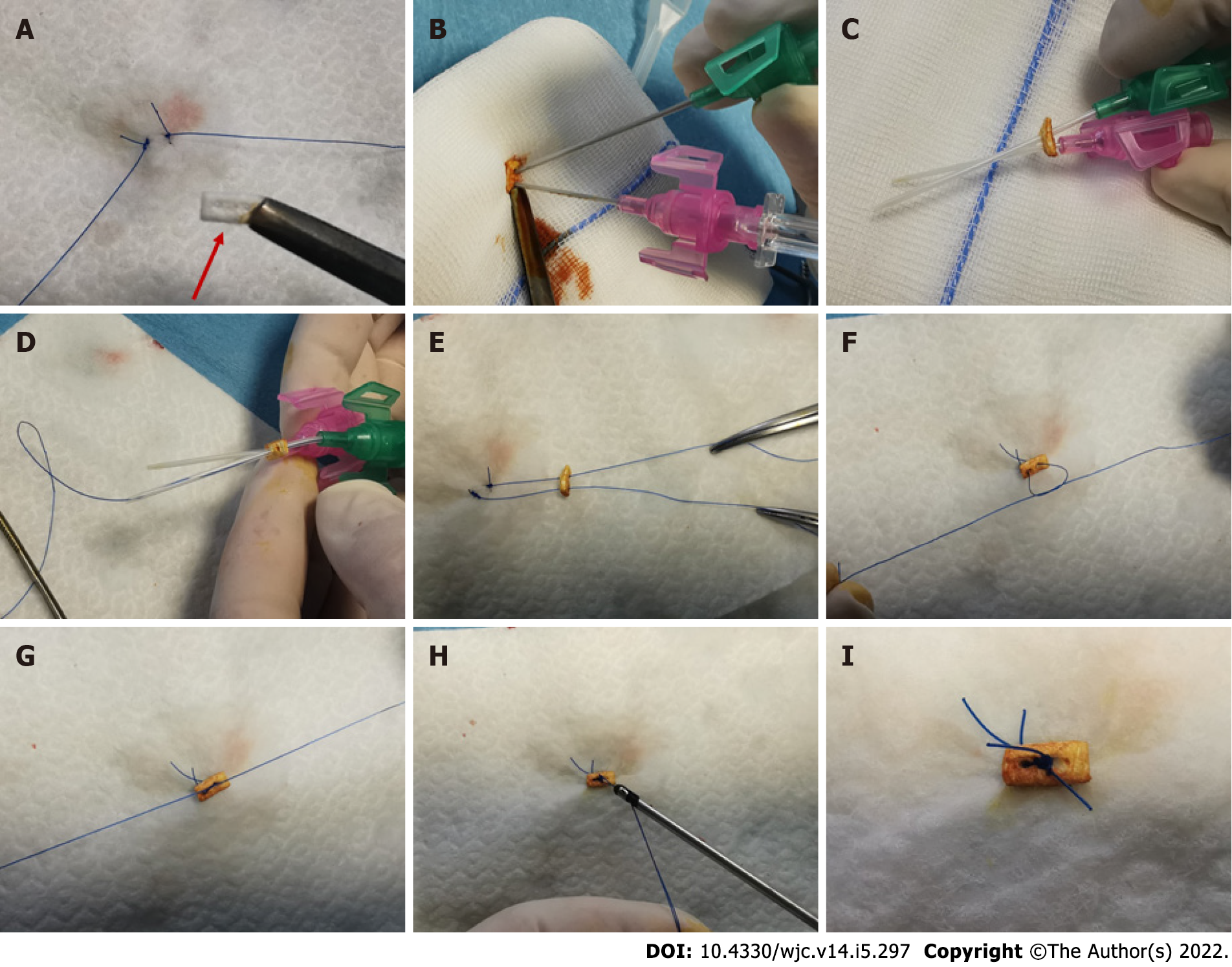
Figure 2 Steps of pledget-assisted hemostasis.
The technique is shown as practiced on a white drape in order to show the steps in the absence of blood. A: Double ProGlide suture after cutting one monofilament from each device and pledget (red arrow); B: Insertion of two cannulas through the pledget (colored by iodine solution to facilitate recognition); C: Steel needles removal from the cannulas; D: Insertion of ProGlide monofilaments through the cannulas; E: Cannulas removal leaving Proglide monofilaments inserted through the pledget; F: Realization of one of two knots; G: Pledget fixation on the artery wall tightening the knots; H: Cut of residual Proglide threads; I: Final configuration achieved with pledged tightened over the two ProGlide’s sutures.
- Citation: Burzotta F, Aurigemma C, Kovacevic M, Romagnoli E, Cangemi S, Bianchini F, Nesta M, Bruno P, Trani C. Pledget-assisted hemostasis to fix residual access-site bleedings after double pre-closure technique. World J Cardiol 2022; 14(5): 297-306
- URL: https://www.wjgnet.com/1949-8462/full/v14/i5/297.htm
- DOI: https://dx.doi.org/10.4330/wjc.v14.i5.297


