Copyright
©The Author(s) 2022.
World J Cardiol. Apr 26, 2022; 14(4): 260-265
Published online Apr 26, 2022. doi: 10.4330/wjc.v14.i4.260
Published online Apr 26, 2022. doi: 10.4330/wjc.v14.i4.260
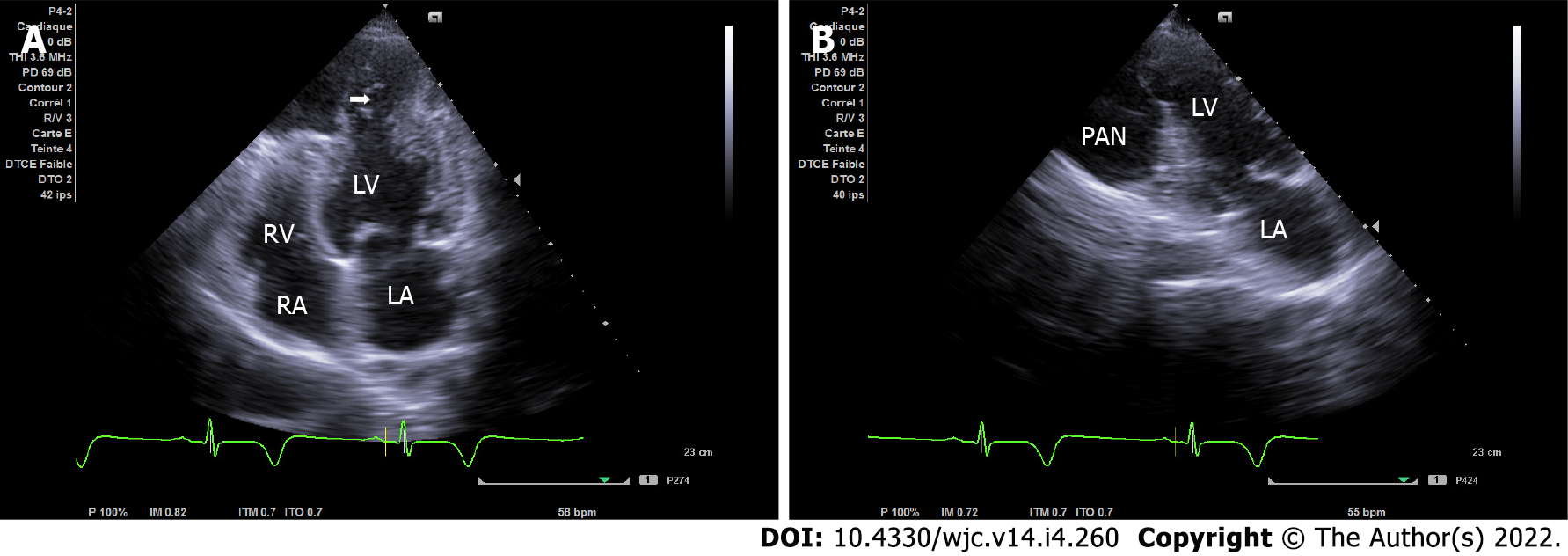
Figure 1 Apical chamber view on transthoracic echocardiogram.
A: Apical four-chamber view demonstrating the narrow neck of a pseudoaneurysm (PAN) in the apical wall (arrow indicates the site of free wall rupture); B: Apical two-chamber view demonstrating a second PAN of the infero-apical wall. LA: Left atrium; LV: Left ventricular; RA: Right atrium; RV: Right ventricular.
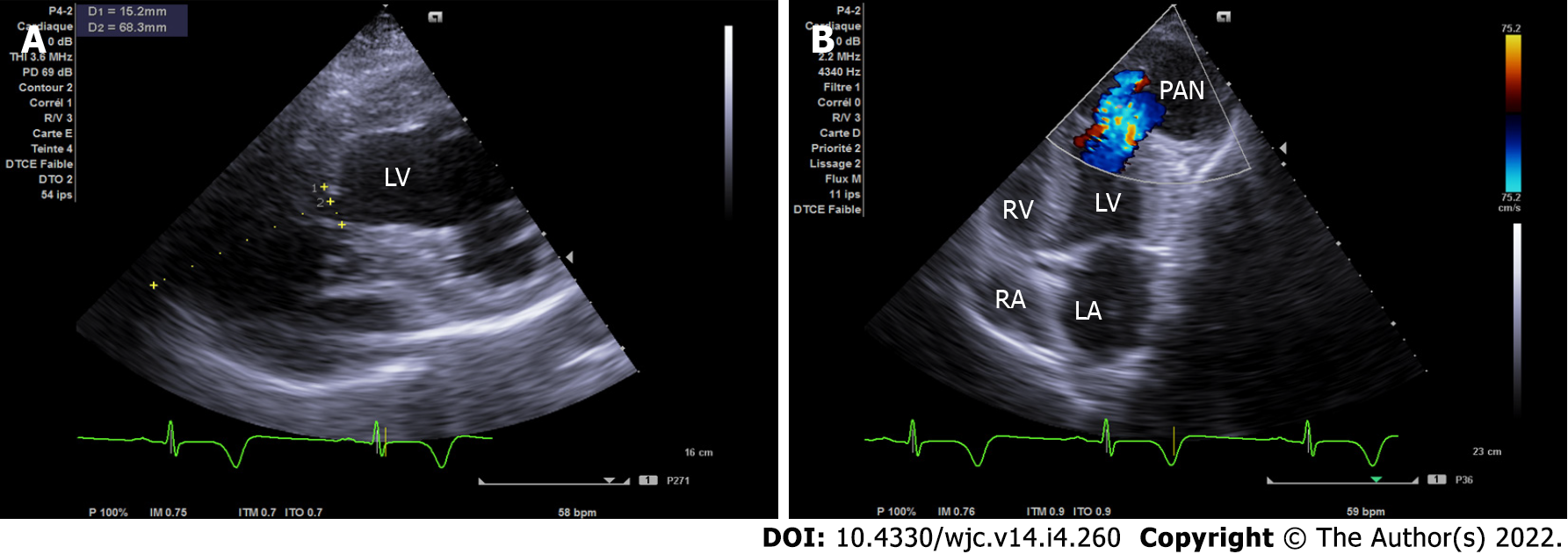
Figure 2 Transthoracic echocardiogram.
A: Modified short axis view with the pseudoaneurysm dimensions (width of the neck was 15 mm and the maximal internal diameter of the aneurysmal sac was 68 mm with a neck to sac ratio of less than 0.5); B: Apical modified 4/5 chamber view showing bidirectional shunt through the left ventricular wall.
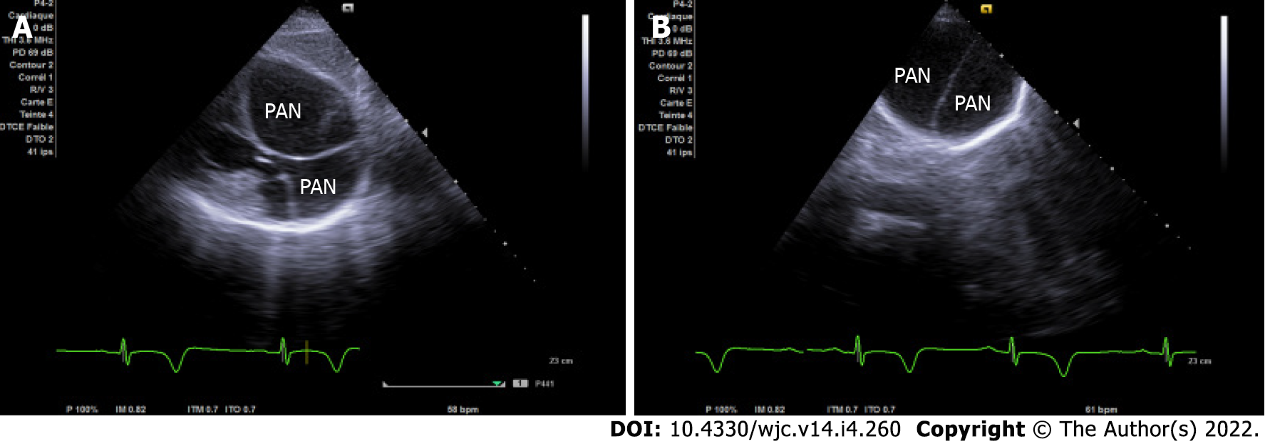
Figure 3 Subcostal and apical views on transthoracic echocardiogram.
A: Subcostal view illustrating two wide pseudoaneurysms isolated by organizing fibrous tissue; B: Apical view with inclination of the probe at a right angle to the skin showing two non-communicant adjacent cavities separated by fibrous tissue.
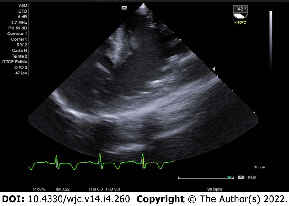
Figure 4 Two-chamber view on transesophageal echocardiogram.
Two-chamber view confirming the pseudoaneurysm and flow through the left ventricle.
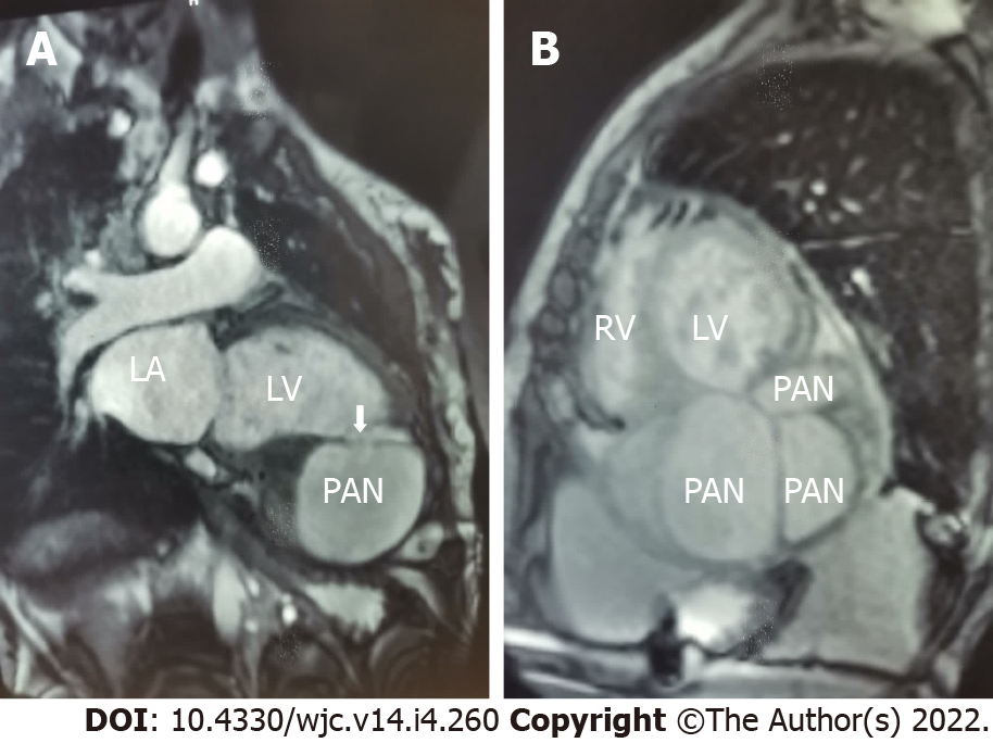
Figure 5 Cardiac magnetic resonance imaging.
A: Two-chamber view showing the connection between the left ventricular (LV) cavity and the LV pseudoaneurysm (PAN) (arrow); B: Short axis view showing multiple cavities corresponding to the PANs isolated by fibrous tissue. LA: Left atrium; RV: Right ventricular.
- Citation: Jallal H, Belabes S, Khatouri A. Uncommon post-infarction pseudoaneurysms: A case report. World J Cardiol 2022; 14(4): 260-265
- URL: https://www.wjgnet.com/1949-8462/full/v14/i4/260.htm
- DOI: https://dx.doi.org/10.4330/wjc.v14.i4.260


