Copyright
©The Author(s) 2022.
World J Cardiol. Apr 26, 2022; 14(4): 239-249
Published online Apr 26, 2022. doi: 10.4330/wjc.v14.i4.239
Published online Apr 26, 2022. doi: 10.4330/wjc.v14.i4.239
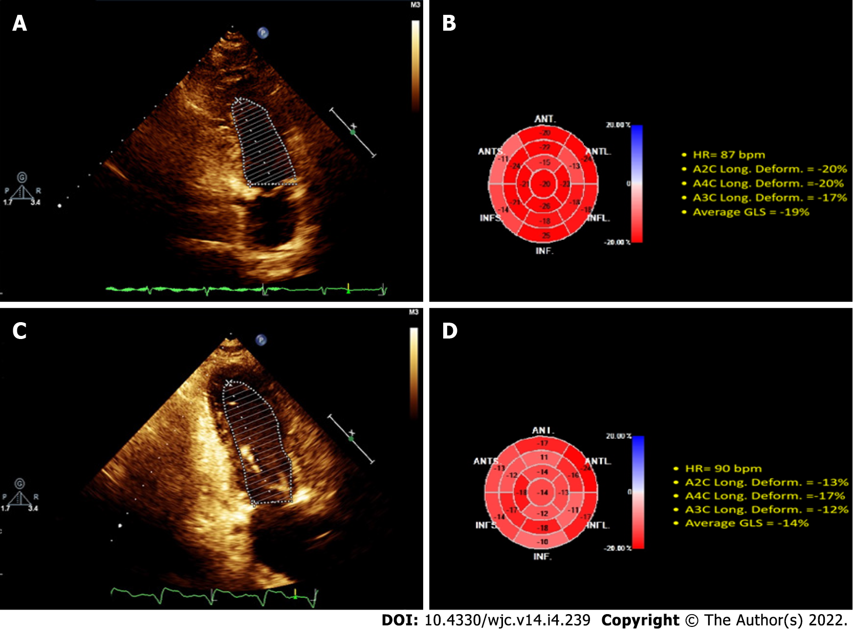
Figure 1 Illustrative 2D echocardiography.
Top: control patient; Bottom: patient with end-stage renal disease (ESRD) and severe hyperparathyroidism (SHPTH). The ejection fraction (EF) by the Simpson method was calculated as a function of the endocardial borders at end-diastole and end-systole in the apical projection of two cavities. A and C (left): in the control patient, left ventricular EF (LVEF) was 56% (A), and 61% in the patient with ESRD and SHPTH (C). 2D ECHO ST real-time showed longitudinal deformation (B and D, right). The deformation pattern in the control patient (B) was normal, with global longitudinal strain (GLS) = 19%, and GLS was abnormal (14%) in the patient with ESRD and SHPTH (D).
- Citation: Carrasco-Ruiz MF, Ruiz-Rivera A, Soriano-Ursúa MA, Martinez-Hernandez C, Manuel-Apolinar L, Castillo-Hernandez C, Guevara-Balcazar G, Farfán-García ED, Mejia-Ruiz A, Rubio-Gayosso I, Perez-Capistran T. Global longitudinal strain is superior to ejection fraction for detecting myocardial dysfunction in end-stage renal disease with hyperparathyroidism. World J Cardiol 2022; 14(4): 239-249
- URL: https://www.wjgnet.com/1949-8462/full/v14/i4/239.htm
- DOI: https://dx.doi.org/10.4330/wjc.v14.i4.239


