Copyright
©The Author(s) 2022.
World J Cardiol. Apr 26, 2022; 14(4): 206-219
Published online Apr 26, 2022. doi: 10.4330/wjc.v14.i4.206
Published online Apr 26, 2022. doi: 10.4330/wjc.v14.i4.206
Figure 1 Different locations of myxoma.
A-C: Cardiac myxoma involving the left atrium (A and C) and the right atrium (B); A, B: The myxoma mass is attached to the atrial septum; C: The tumour is related to the lateral wall of left atrium.
Figure 2 Biatrial myxoma in a 22-yr-old Bangladeshi male.
A: 2-D transthoracic echocardiography shows biatrial myxoma arising from the midinteratrial septum; B, C: Tumour mass during and after surgery.
Figure 3 Carney complex in a middle-aged Bangladeshi female.
A: Multiple pigmented lentigines distributed symmetrically on the face of the patient; B: 2-D transthoracic echocardiography shows multichamber myxoma involving the left atrium, left ventricle and the right atrium; C: 2-D transthoracic echocardiography of the son of the lady shows myxoma in the right atrium.
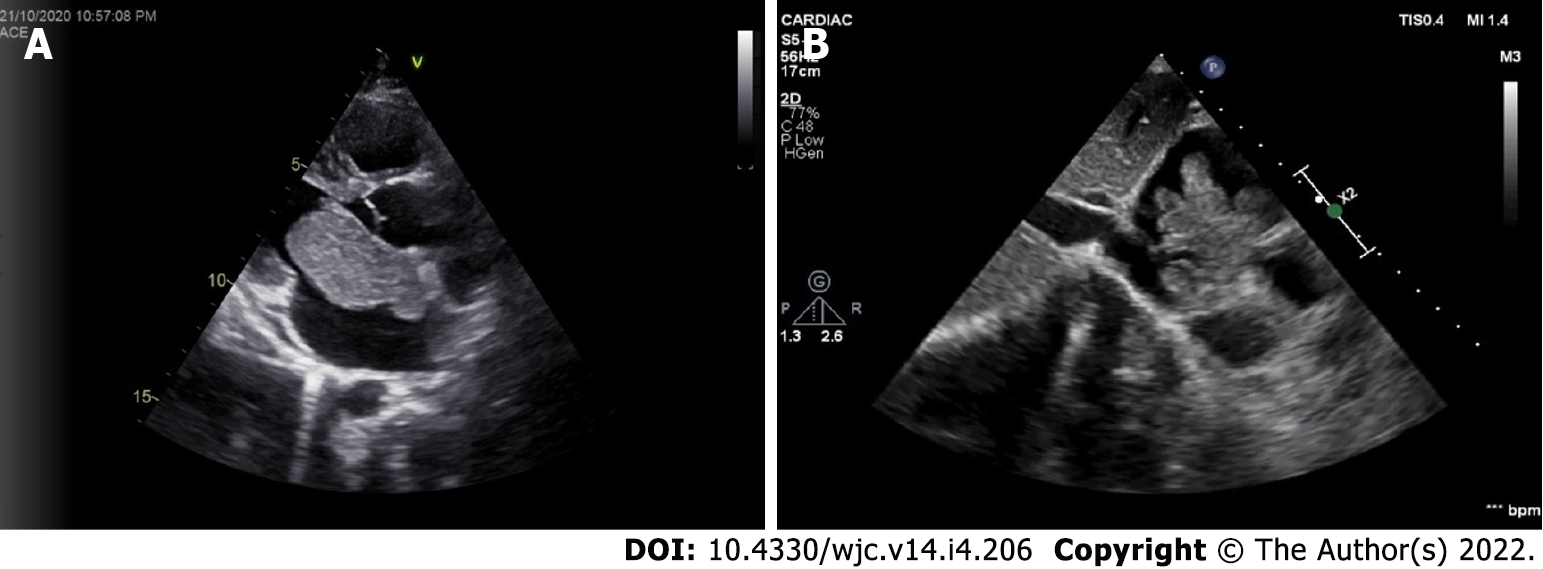
Figure 4 Polypoid and papillary myxoma in 2D echocardiography.
A: Parasternal long axis view shows a polypoid left atrial myxoma; B: Subcostal view shows a papillary right atrial myxoma with multiple projections.
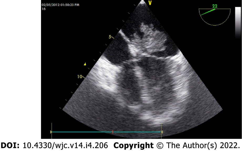
Figure 5 Papillary myxoma presenting with ischemic stroke.
Transesophageal echocardiography shows a fragile papillary myxoma. The young patient presented with acute ischemic stroke.
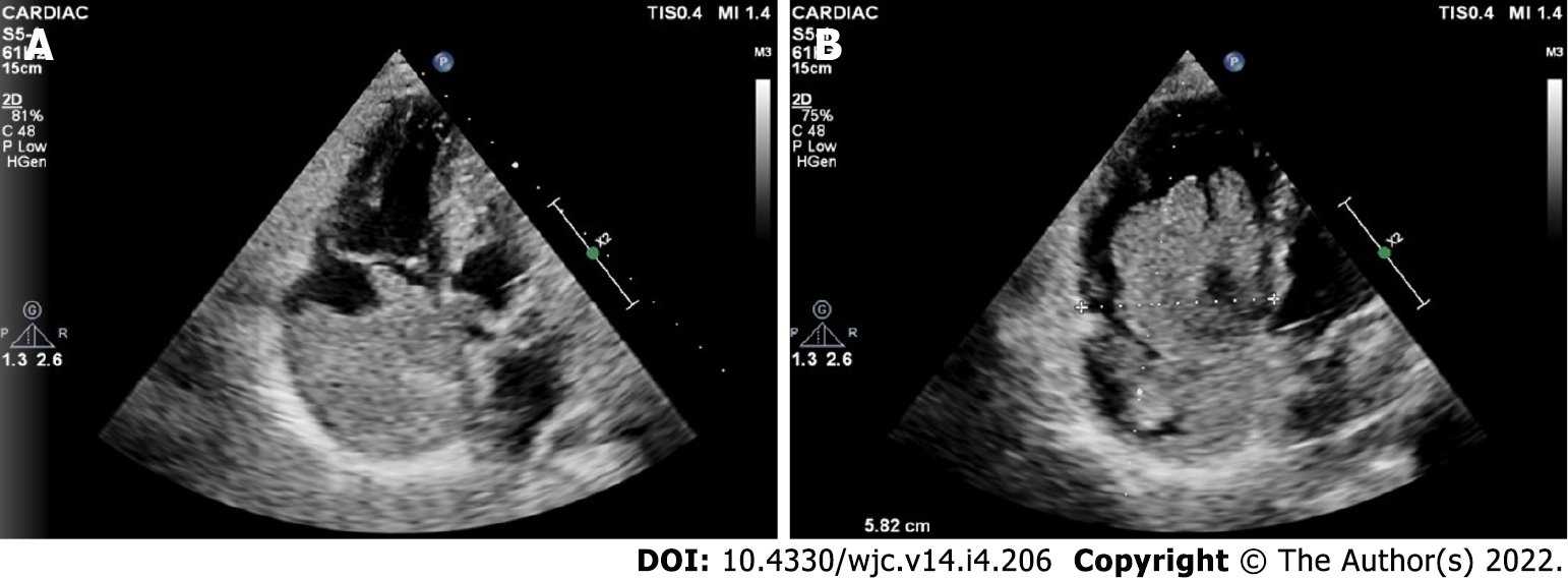
Figure 6 Giant myxoma in right atrium.
A, B: 2D transthoracic echocardiography shows a giant myxoma occupying the major parts of the right atrial cavity. Note the multiple papillary projections evident in (B).
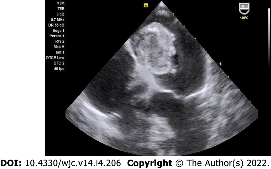
Figure 7 Calcified myxoma in the left atrium.
Transesophageal echocardiography shows a large calcified myxoma occupying the left atrial cavity.
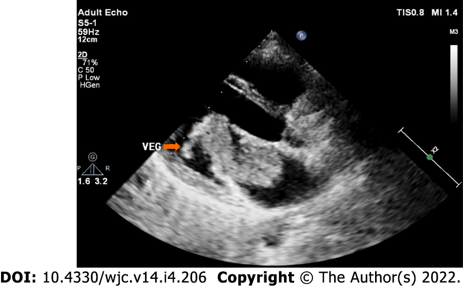
Figure 8 Infected myxoma in the left atrium.
Transthoracic echocardiography shows a large left atrial myxoma protruding into the left ventricular cavity across the mitral valve. A vegetation with independent mobility is attached to the tumour. The patient presented with prolonged fever with positive blood culture.
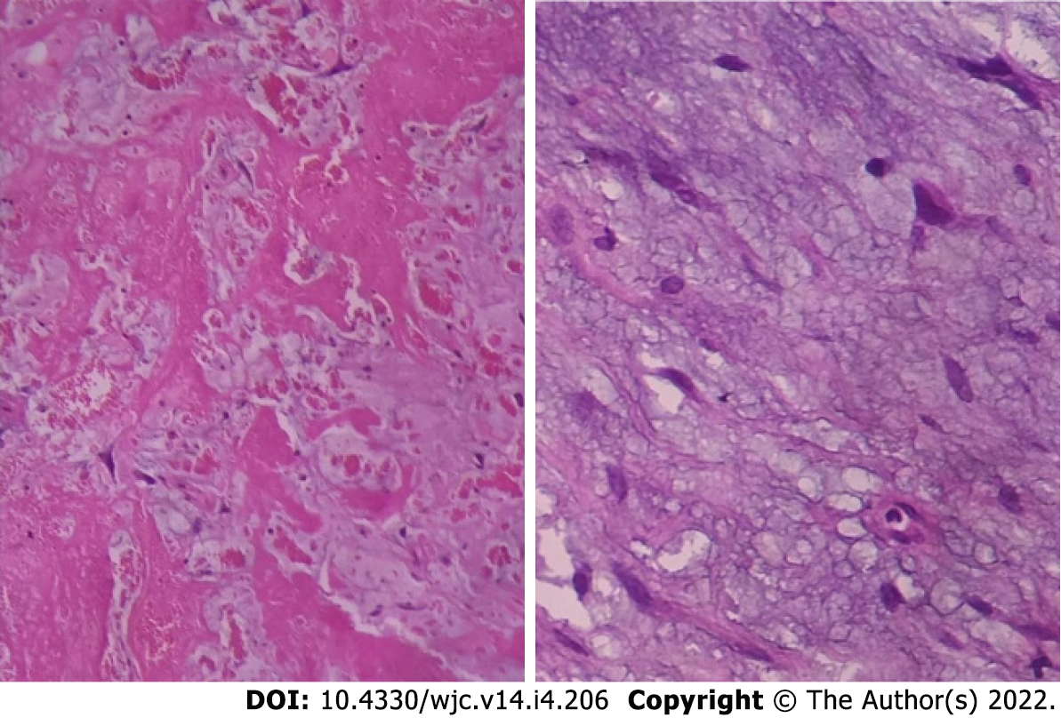
Figure 9 Histopathology of myxoma.
Histopathological examination shows abundant loose myxoid stroma with scattered round, polygonal or stellate cells with dense irregular nuclei.
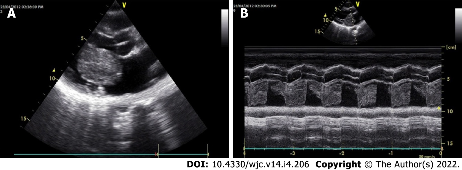
Figure 10 Echocardiographic features of myxoma.
A, B: Transthoracic echocardiography 2D (A) and M-mode (B) shows a large polypoid mass in the left atrial cavity attached to the interatrial septum by means of a stalk (not visualized here) and protruding into the left ventricular cavity across the mitral valve in diastole.
- Citation: Islam AKMM. Cardiac myxomas: A narrative review. World J Cardiol 2022; 14(4): 206-219
- URL: https://www.wjgnet.com/1949-8462/full/v14/i4/206.htm
- DOI: https://dx.doi.org/10.4330/wjc.v14.i4.206


