Copyright
©The Author(s) 2022.
World J Cardiol. Apr 26, 2022; 14(4): 190-205
Published online Apr 26, 2022. doi: 10.4330/wjc.v14.i4.190
Published online Apr 26, 2022. doi: 10.4330/wjc.v14.i4.190
Figure 1 Cardiac magnetic resonance imaging of acute myocardial infarction.
A: Short axis mid-ventricular image demonstrating almost full-thickness transmural late gadolinium enhancement (LGE) in posterolateral wall (yellow arrow); B: Four-chamber image demonstrating focal LGE in lateral wall (red arrow); C: Short axis image demonstrating > 75% transmural LGE in lateral wall (orange arrow).
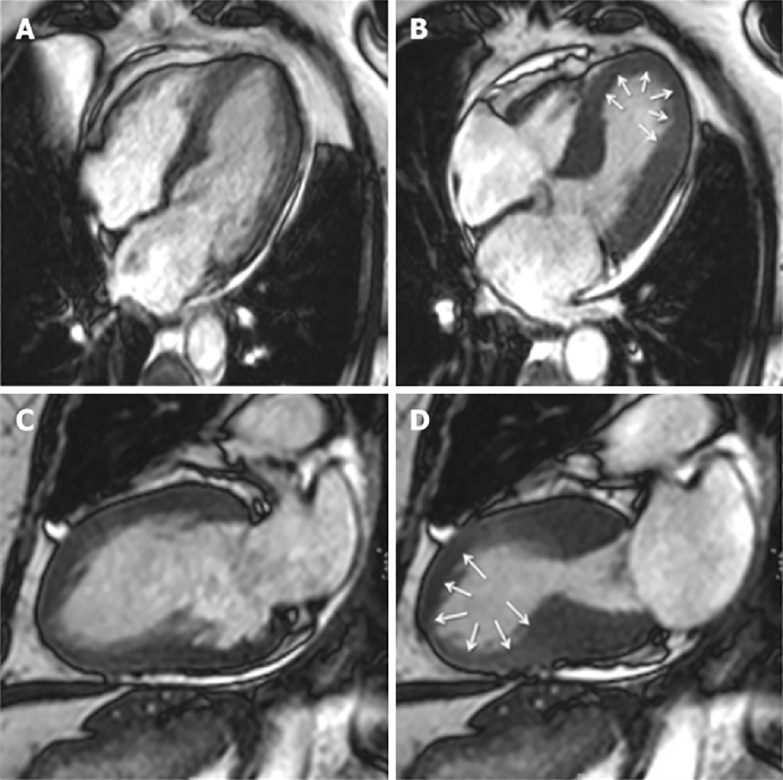
Figure 2 Cardiac magnetic resonance imaging of Takotsubo cardiomyopathy.
Typical apical ballooning seen in takotsubo syndrome. A, B: Cine four-chamber in late diastole and systole respectively; C, D: Two-chamber view in late diastole and systole respectively. Modified from Plácido et al[123] and licensed under the Creative Commons Attribution 4.0 International License (http://creativecommons.org/licenses/by/4.0/).
Figure 3 Cardiac magnetic resonance imaging of acute myocarditis.
A: Four-chamber image demonstrating LGE in septal wall in a mid-wall pattern (yellow arrow); B: Short axis mid-left ventricular image demonstrating LGE in anteroseptal wall in a mid-wall pattern (red arrow); C: Two-chamber image demonstrating LGE in the anterior wall in a mid-wall pattern (orange yellow).
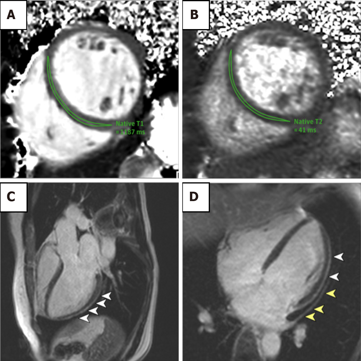
Figure 4 COVID-19 related cardiac dysfunction on cardiac magnetic resonance imaging.
Cardiac magnetic resonance imaging of an adult woman with COVID-19-related perimyocarditis. A, B: Significantly raised native T1 and native T2 in myocardial mapping acquisitions; C, D: Pericardial effusion and enhancement (yellow arrowheads) and epicardial and intramyocardial enhancement (white arrowheads) using LGE acquisition. Modified from Puntmann et al[75] and licensed under CC BY 4.0 (https://creativecommons.org/licenses/by/4.0/).
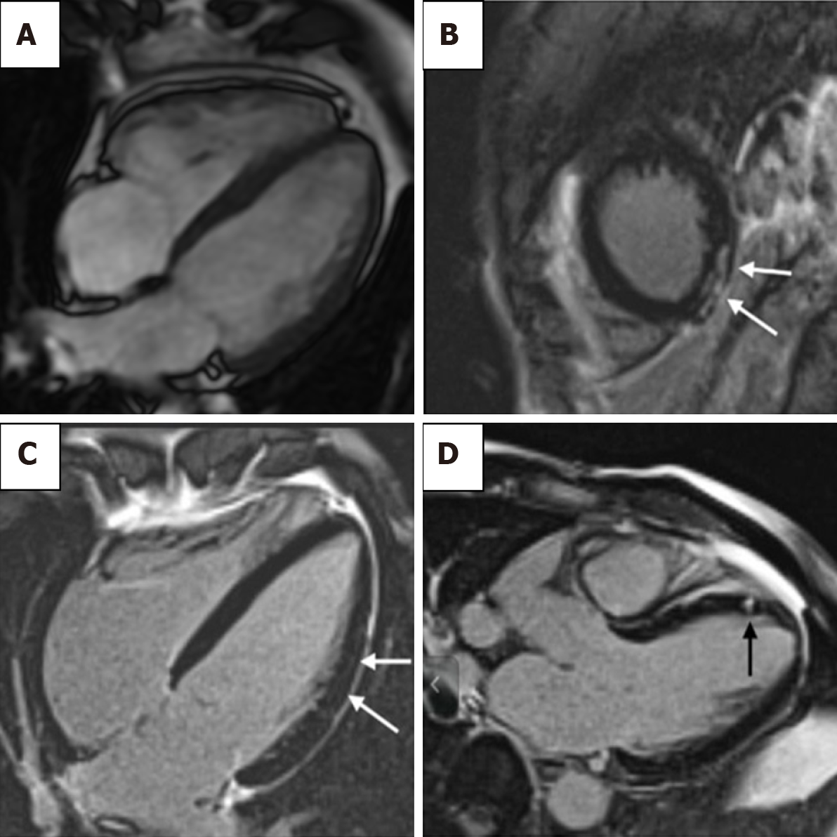
Figure 5 Cardiac magnetic resonance imaging of athlete’s heart syndrome.
A: Cardiac magnetic resonance imaging of an endurance athlete. Increased right and left ventricular volumes. Overall muscle mass may be increased although wall thickness remains within standard reference range[102]; B: A 51-year-old athlete training 7 h/wk in the last 30 years. The short-axis view shows subepicardial late gadolinium enhancement (LGE) in the inferior apical wall; C: A 55-year-old athlete training 8 h/wk in the last 30 years. Mild intramyocardial LGE is the lateral wall is shown in the four-chamber view; D: A 55-year-old athlete training 10 h/wk in the last 28 years. Mesocardial LGE in the apical-septal wall shown in three-chamber view image. Reproduced from Pujadas et al[122] and licensed under CC BY 4.0 (https://creativecommons.org/licenses/by/4.0/).
- Citation: Nguyen Nguyen N, Assad JG, Femia G, Schuster A, Otton J, Nguyen TL. Role of cardiac magnetic resonance imaging in troponinemia syndromes. World J Cardiol 2022; 14(4): 190-205
- URL: https://www.wjgnet.com/1949-8462/full/v14/i4/190.htm
- DOI: https://dx.doi.org/10.4330/wjc.v14.i4.190


