Copyright
©The Author(s) 2022.
World J Cardiol. Oct 26, 2022; 14(10): 546-556
Published online Oct 26, 2022. doi: 10.4330/wjc.v14.i10.546
Published online Oct 26, 2022. doi: 10.4330/wjc.v14.i10.546
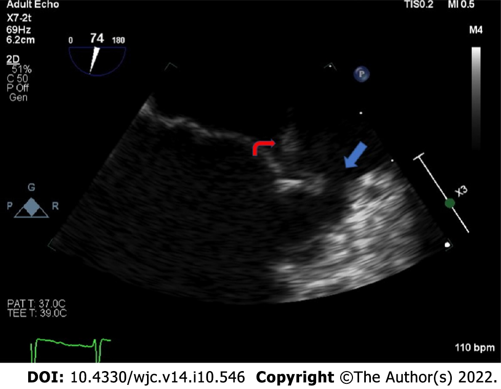
Figure 1 Zoomed mid-esophageal view on transesophageal echocardiography, showing an echodensity attached to the A2 segment of the anterior leaflet of the mitral valve (red arrow) with evidence of leaflet perforation in the A1 segment (blue arrow).
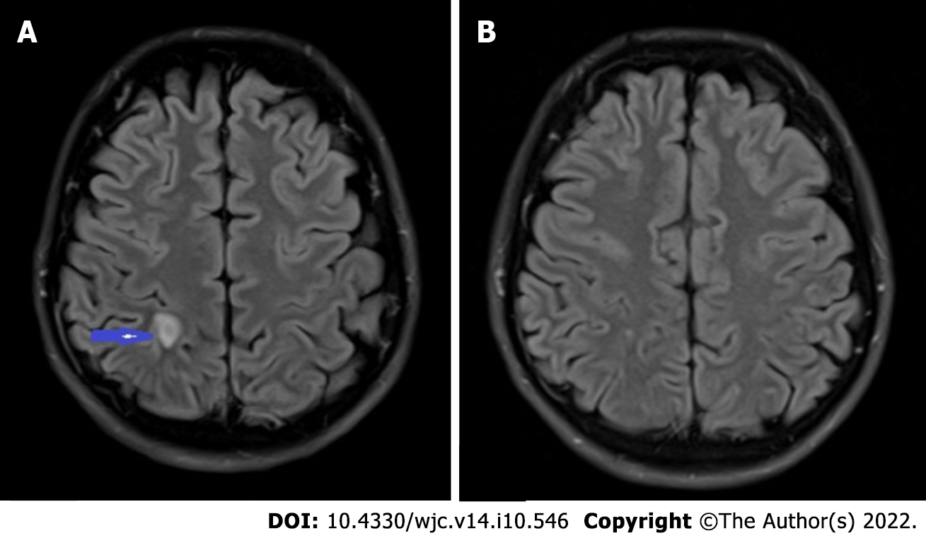
Figure 2 Septic embolus in the brain.
A: T2-weighted MRI brain showing a 1.0 cm × 0.5 cm ring enhancing lesion (blue arrow) in the right parietal lobe with central diffusion restriction and mild surrounding vasogenic edema; B: Repeat MRI brain with near complete resolution of the ring enhancing lesion. MRI: Magnetic resonance imaging.
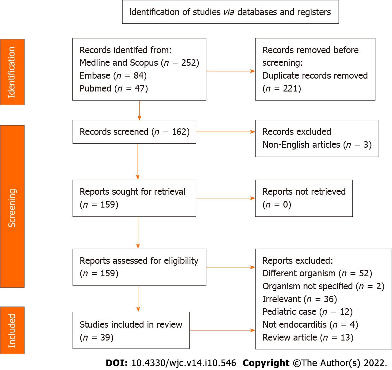
Figure 3 PRISMA flow chart highlighting article search and selection.
- Citation: Olagunju A, Martinez J, Kenny D, Gideon P, Mookadam F, Unzek S. Virulent endocarditis due to Haemophilus parainfluenzae: A systematic review of the literature. World J Cardiol 2022; 14(10): 546-556
- URL: https://www.wjgnet.com/1949-8462/full/v14/i10/546.htm
- DOI: https://dx.doi.org/10.4330/wjc.v14.i10.546


