Copyright
©The Author(s) 2022.
World J Cardiol. Oct 26, 2022; 14(10): 522-536
Published online Oct 26, 2022. doi: 10.4330/wjc.v14.i10.522
Published online Oct 26, 2022. doi: 10.4330/wjc.v14.i10.522
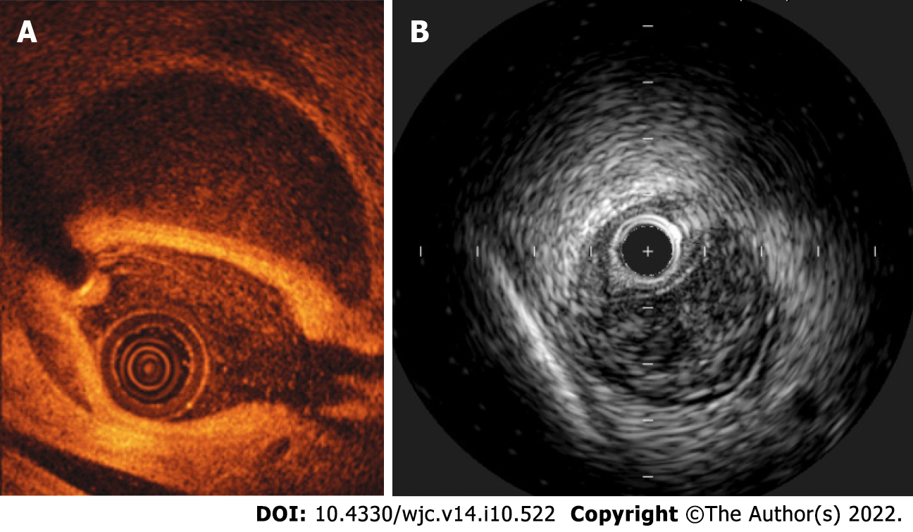
Figure 1 Intravascular imaging techniques in spontaneous coronary artery dissection.
A: Optical coherence tomography; B: Intravascular ultrasound.
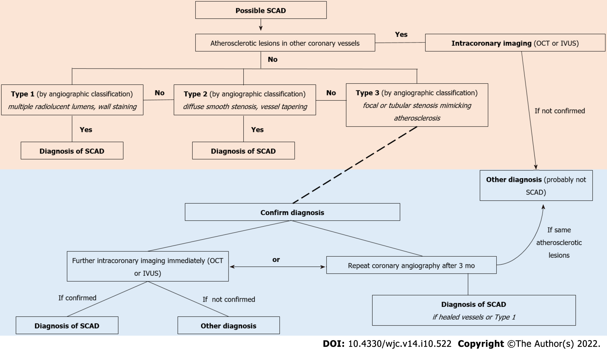
Figure 2 Flowchart diagram for the diagnosis of spontaneous coronary artery dissection.
OCT: Optical coherence tomography; IVUS: Intravascular ultrasound; SCAD: Spontaneous coronary artery dissection.
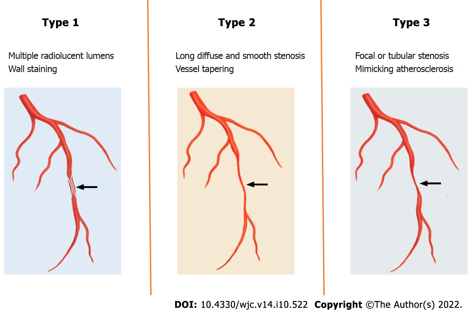
Figure 3 Types of spontaneous coronary artery dissection based on angiographic imaging techniques.
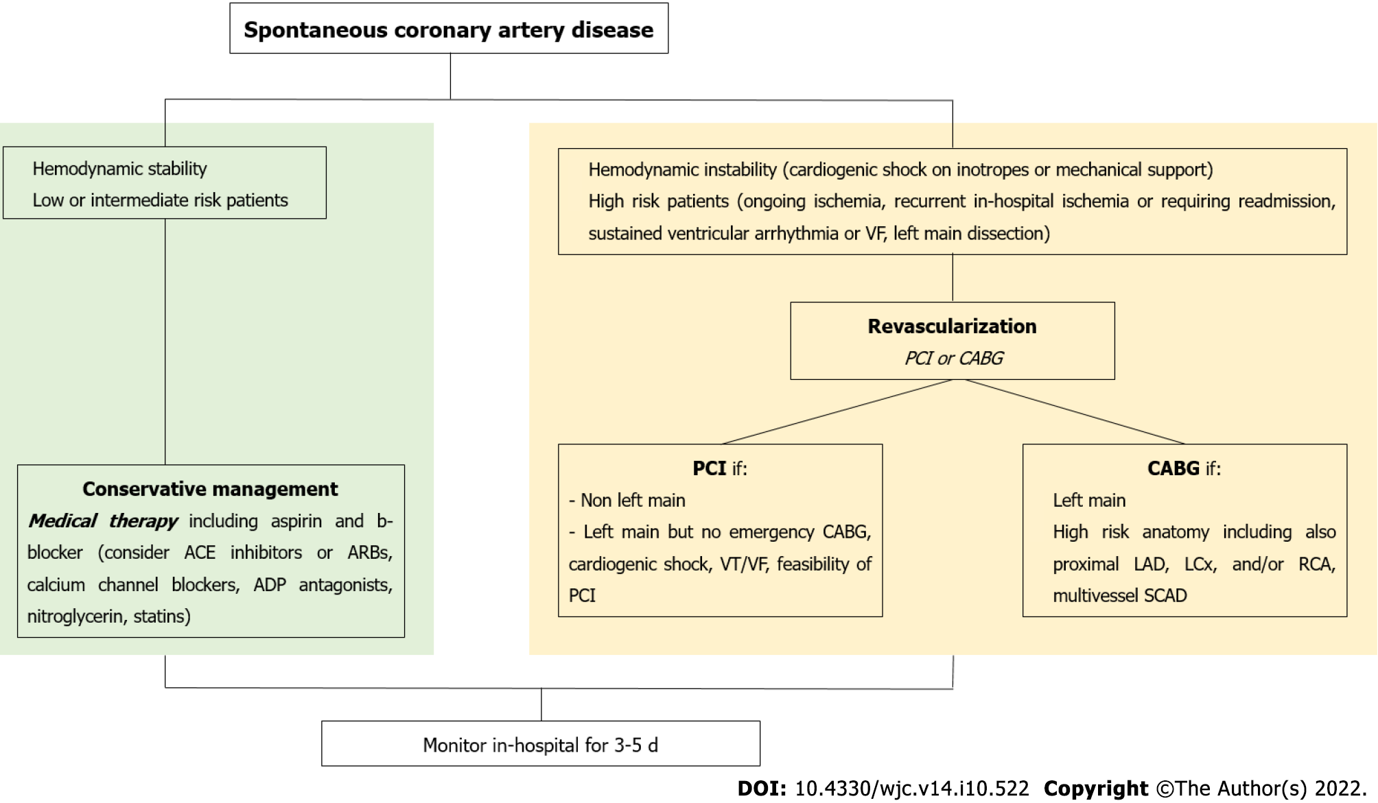
Figure 4 Proposed management algorithm for spontaneous coronary artery dissection.
VF: Ventricular fibrillation; ACE: Angiotensin converting enzyme; ADP: Adenosine diphosphate; CABG: Coronary artery by-pass grafting; VT: Ventricular tachycardia; PCI: Percutaneous coronary intervention; LAD: Left anterior descending artery; LCx: Left circumflex coronary; RCA: Right coronary artery; SCAD: Spontaneous coronary artery dissection.
- Citation: Lionakis N, Briasoulis A, Zouganeli V, Dimopoulos S, Kalpakos D, Kourek C. Spontaneous coronary artery dissection: A review of diagnostic methods and management strategies. World J Cardiol 2022; 14(10): 522-536
- URL: https://www.wjgnet.com/1949-8462/full/v14/i10/522.htm
- DOI: https://dx.doi.org/10.4330/wjc.v14.i10.522


