Copyright
©The Author(s) 2022.
World J Cardiol. Jan 26, 2022; 14(1): 54-63
Published online Jan 26, 2022. doi: 10.4330/wjc.v14.i1.54
Published online Jan 26, 2022. doi: 10.4330/wjc.v14.i1.54
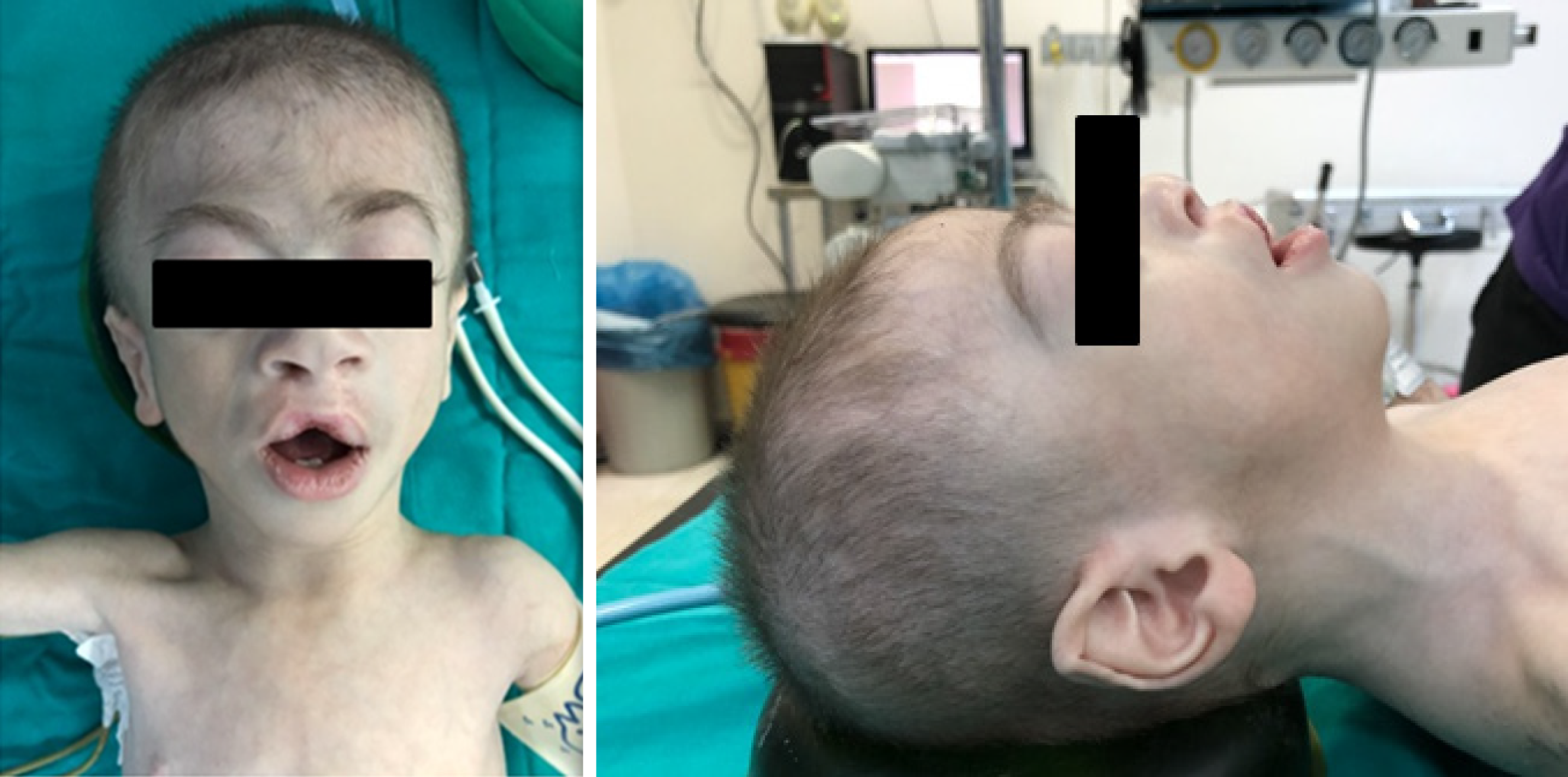
Figure 1 Distinguishing craniofacial features of the patient.
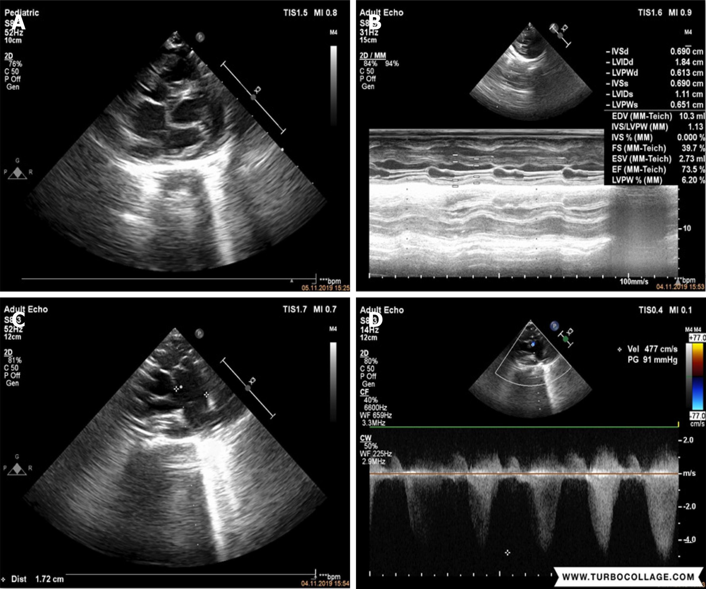
Figure 2 Echocardiographic images.
A: The heart of the patient; B: Mild to moderate regurgitation in the tricuspid valve; C: Left ventricular systolic function was observed, functions were maintained (EF: 73.5 %, FS: 39.7 %); D: 91 mmHg gradient was obtained at pulmonary valve level in parasternal short axis imaging.
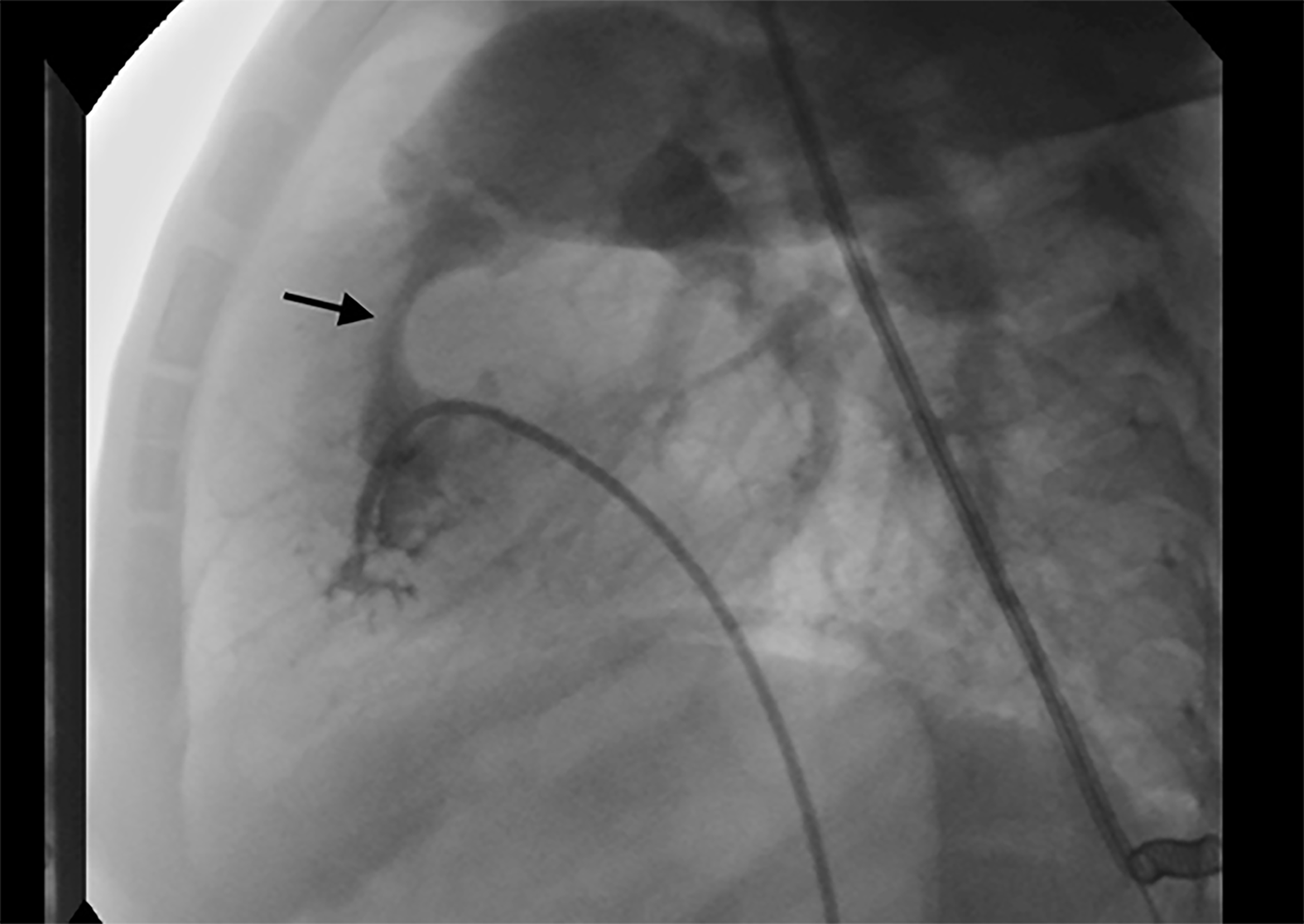
Figure 3 Appearance of the stenotic pulmonary valve in the angiography image.
- Citation: Arun O, Oc B, Metin EN, Sert A, Yilmaz R, Oc M. Anesthetic management of a child with Cornelia de Lange Syndrome undergoing open heart surgery: A case report. World J Cardiol 2022; 14(1): 54-63
- URL: https://www.wjgnet.com/1949-8462/full/v14/i1/54.htm
- DOI: https://dx.doi.org/10.4330/wjc.v14.i1.54


