Copyright
©The Author(s) 2021.
World J Cardiol. Apr 26, 2021; 13(4): 111-116
Published online Apr 26, 2021. doi: 10.4330/wjc.v13.i4.111
Published online Apr 26, 2021. doi: 10.4330/wjc.v13.i4.111
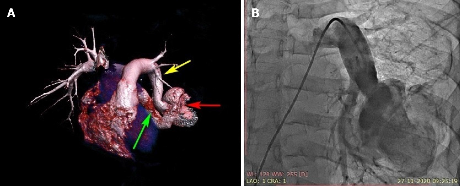
Figure 1 Three-dimensional computed tomography volume rendering and pulmonary artery angiogram.
A: Contrast-enhanced computed tomography with three-dimensional reconstruction showing the left lower pulmonary artery-to-left atrial fistula with a highly tortuous course associated with an aneurysmal sac. Yellow arrow: left lower segmental artery; red arrow: aneurysmal sac; green arrow: last part of fistula connecting the left atrium; B: Anterior-posterior projection of pulmonary artery angiography using a 6-Fr pigtail catheter showing a highly tortuous large left pulmonary artery-to-left atrial fistula associated with a 6-cm aneurysmal sac. The right ventricular oxygen saturation was 79%, and the left ventricular saturation was 92% in room air without anesthesia.
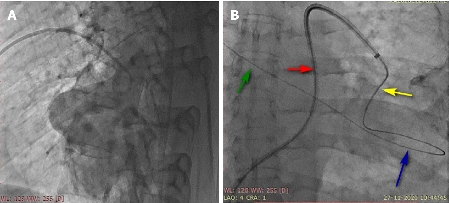
Figure 2 Lateral pulmonary angiography and the course of the guidewire.
A: Lateral pulmonary artery angiography using a 6-Fr pigtail catheter showing a large left pulmonary artery-to-left atrial fistula associated with a large aneurysmal sac; B: A Terumo wire (0.35 cm × 260 cm) and 5-Fr multipurpose catheter helped to negotiate the highly tortuous course of the fistula to reach beyond the targeted landing zone and the site of guide wire placement in the upper right pulmonary vein via the aneurysmal sac for adequate support. Red arrow: 8-Fr sheath; yellow arrow: 5-Fr multipurpose catheter through the delivery sheath; blue arrow: guidewire in the aneurysmal sac; green arrow: upper right pulmonary vein.
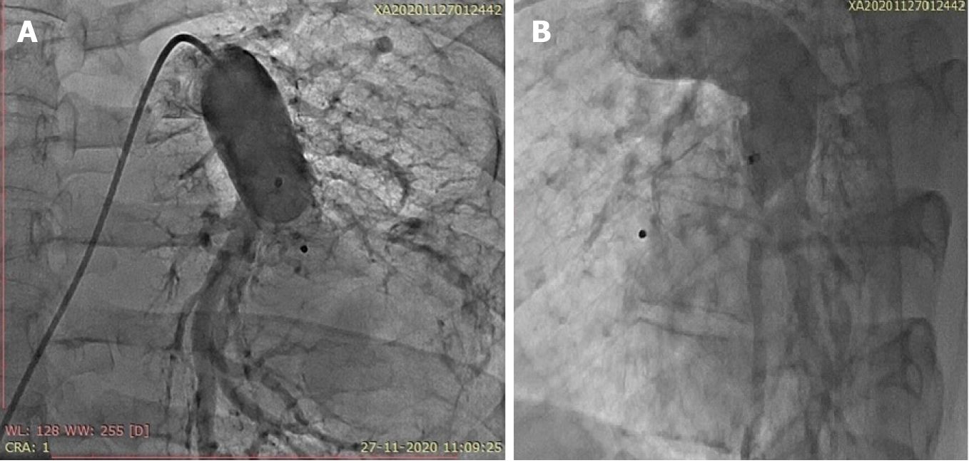
Figure 3 Pulmonary artery angiography after device deployment.
Pulmonary artery angiography in the frontal projection and in the lateral projection showing perfect device positioning, no residual shunting and uncompromised blood flow in the left lower segmental artery. A: Frontal projection; B: Lateral projection.
- Citation: Mahapatra R, Mahanta D, Singh J, Acharya D, Barik R. Device closure of fistula from left lower pulmonary artery to left atrium using a vascular plug: A case report. World J Cardiol 2021; 13(4): 111-116
- URL: https://www.wjgnet.com/1949-8462/full/v13/i4/111.htm
- DOI: https://dx.doi.org/10.4330/wjc.v13.i4.111


