Copyright
©The Author(s) 2021.
World J Cardiol. Jan 26, 2021; 13(1): 28-37
Published online Jan 26, 2021. doi: 10.4330/wjc.v13.i1.28
Published online Jan 26, 2021. doi: 10.4330/wjc.v13.i1.28
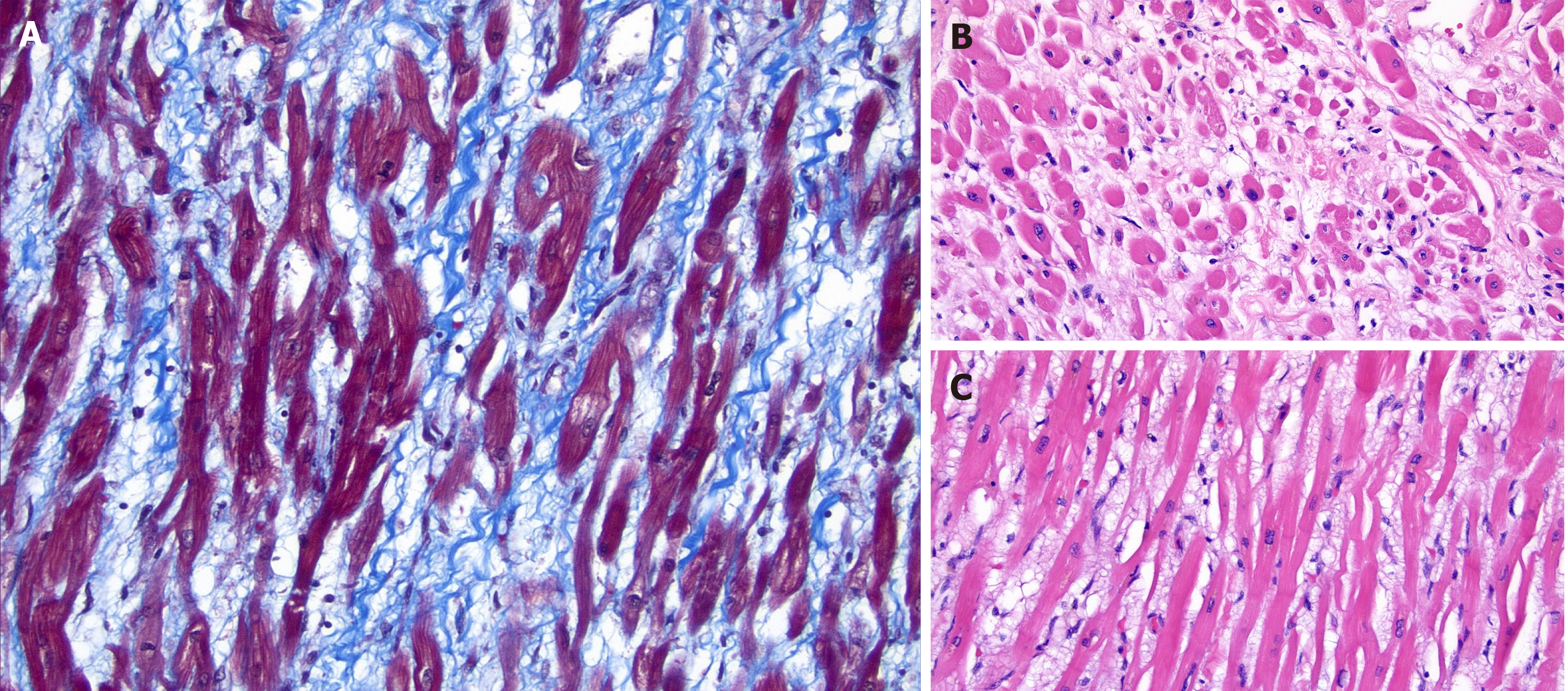
Figure 1 Pathology of myocardium with various stains.
A: Trichrome stain showing minimal collagen deposition; B: Cross section of myocardium showing decreased diameter and prominent interstitial edema; C: Hematoxylin and eosin stain showing mildly thinned myocardiocytes with prominent interstitial edema.
- Citation: Chong EG, Lee EH, Sail R, Denham L, Nagaraj G, Hsueh CT. Anthracycline-induced cardiotoxicity: A case report and review of literature. World J Cardiol 2021; 13(1): 28-37
- URL: https://www.wjgnet.com/1949-8462/full/v13/i1/28.htm
- DOI: https://dx.doi.org/10.4330/wjc.v13.i1.28


