Copyright
©The Author(s) 2020.
World J Cardiol. Jun 26, 2020; 12(6): 285-290
Published online Jun 26, 2020. doi: 10.4330/wjc.v12.i6.285
Published online Jun 26, 2020. doi: 10.4330/wjc.v12.i6.285
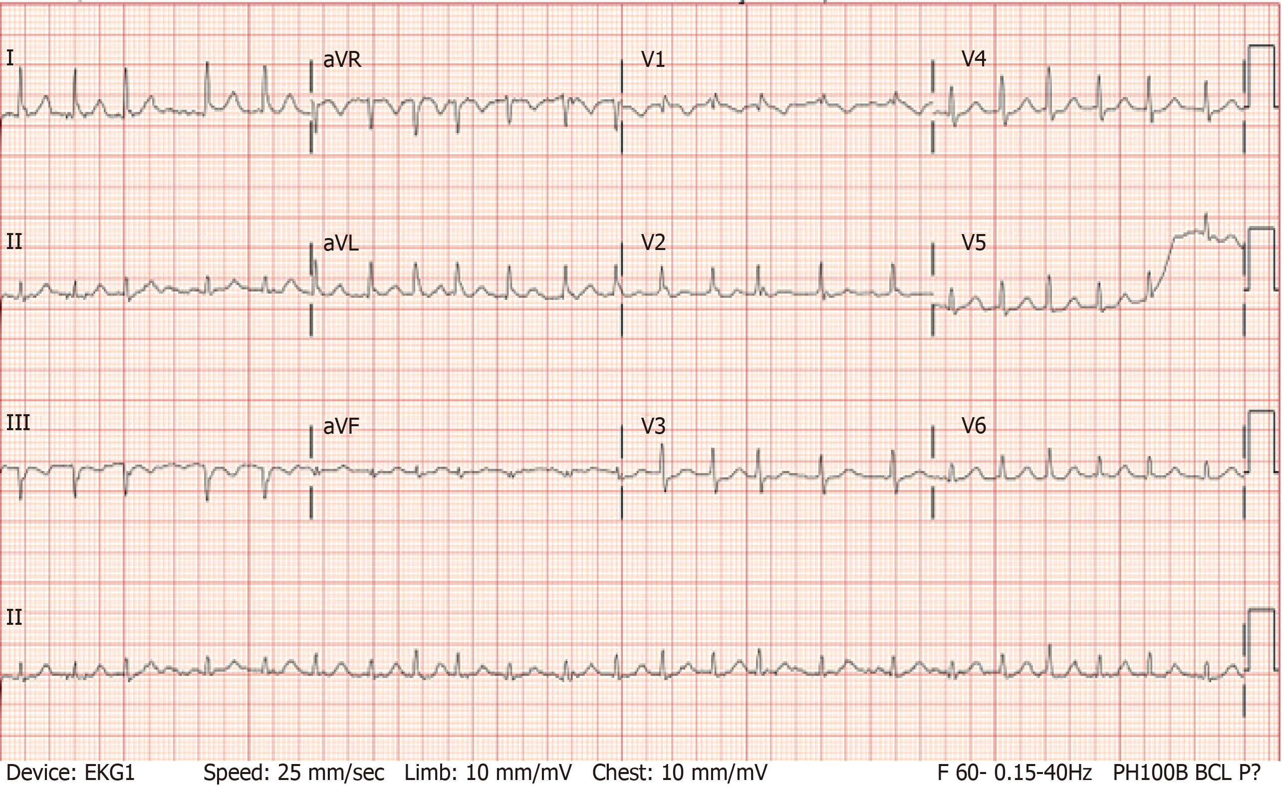
Figure 1 Electrocardiogram showing atrial fibrillation with rapid ventricular response.
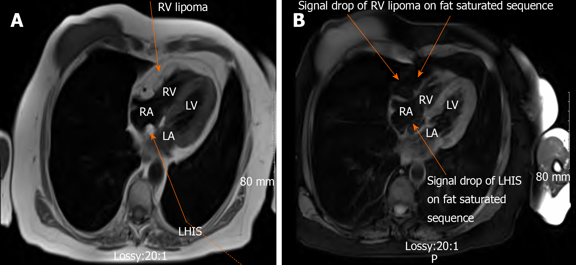
Figure 2 Cardiac magnetic resonance imaging.
A: Axial section T1 weighted imaging showing (a) well-defined capsular homogenous mass along the epicardial surface of right ventricular, diffusely infiltrating the myocardium without frank invasion of adjacent structures (b) lipomatous hypertrophy of interatrial septum; B: Axial section T1 weighted imaging showing signal drop on fat saturated sequence of cardiac lipoma in right ventricular. RV: Right ventricular; LHIS: Lipomatous hypertrophy of interatrial septum.
- Citation: Nalluru SS, Nadadur S, Trivedi N, Trivedi S, Goyal S. Tale of fat and fib — cardiac lipoma managed with radiofrequency ablation: A case report. World J Cardiol 2020; 12(6): 285-290
- URL: https://www.wjgnet.com/1949-8462/full/v12/i6/285.htm
- DOI: https://dx.doi.org/10.4330/wjc.v12.i6.285


