Copyright
©The Author(s) 2020.
World J Cardiol. May 26, 2020; 12(5): 167-191
Published online May 26, 2020. doi: 10.4330/wjc.v12.i5.167
Published online May 26, 2020. doi: 10.4330/wjc.v12.i5.167
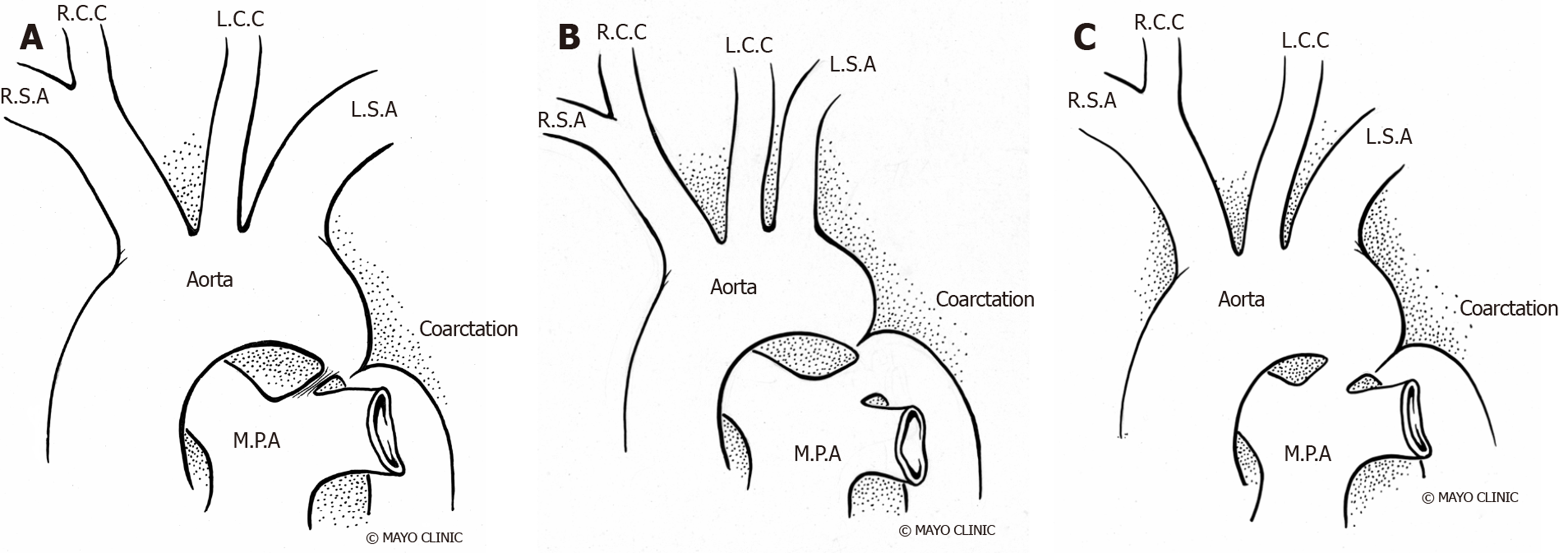
Figure 1 Coarctation of Aorta types.
A: Ductal; B: Pre-ductal; C: Post-ductal. R.S.A: Right subclavian artery; R.C.C: Right common carotid; L.C.C: Left common carotid; L.S.A: Left subclavian artery; M.P.A: Main pulmonary artery.
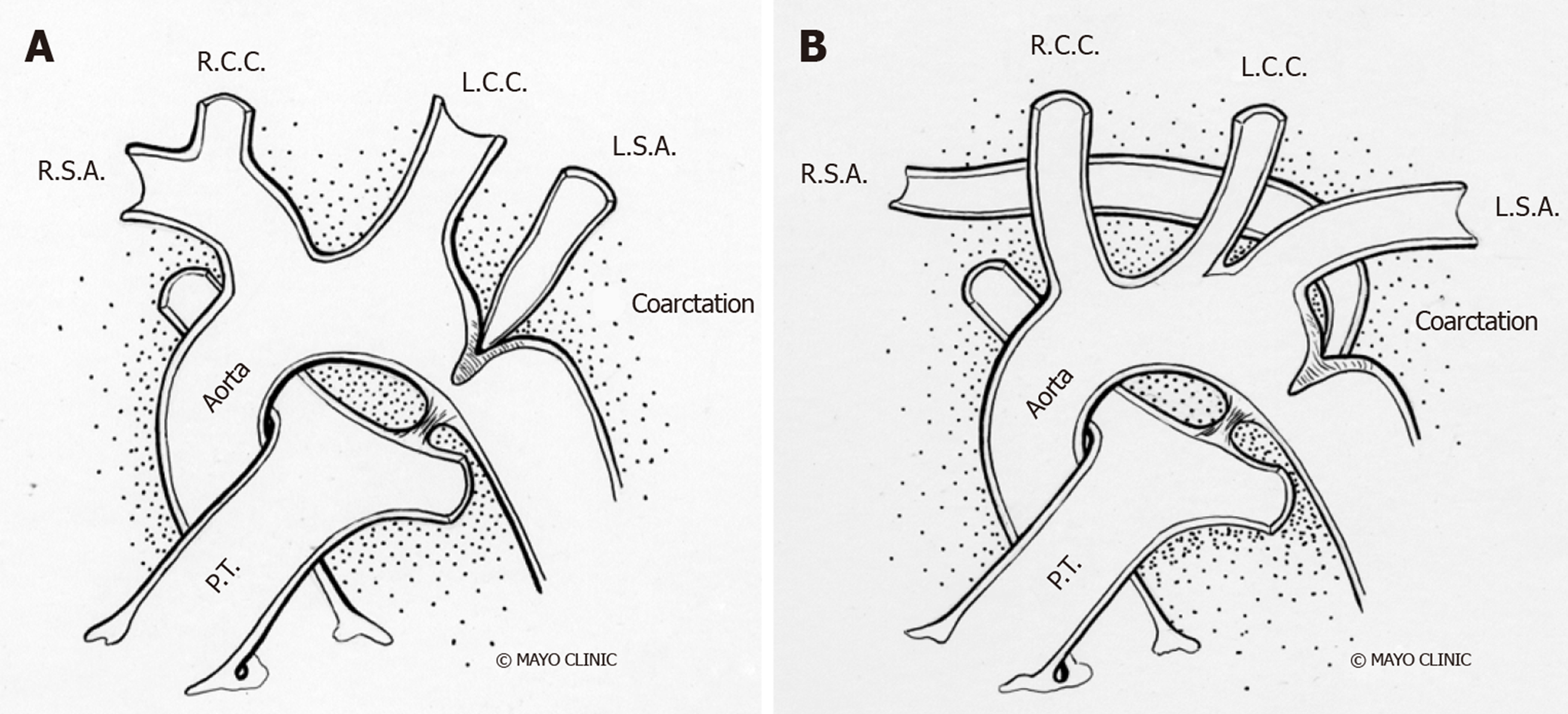
Figure 2 Anatomical variations of subclavian artery in coarctation of aorta.
A: Right subclavian artery arising from the site of coarctation; B: Left subclavian artery arising from the site of coarctation. R.S.A: Right subclavian artery; R.C.C: Right common carotid; L.C.C: Left common carotid; L.S.A: Left subclavian artery; P.T: Pulmonary trunk.
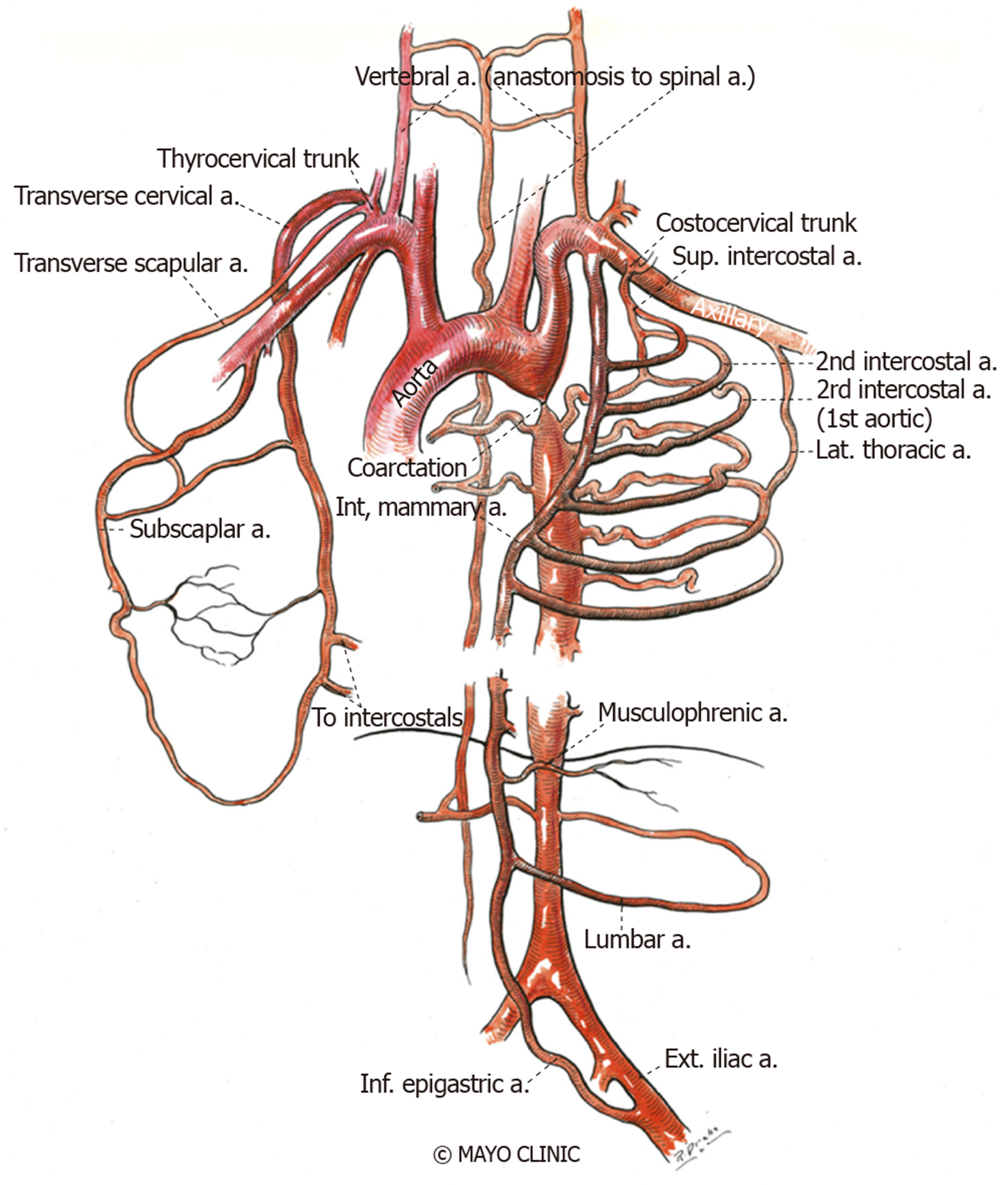
Figure 3 Collateral vessels development due to luminal narrowing, allowing blood flow from high to low pressure areas.
Figure shows the collaterals in intercostal, internal mammary, scapular and lumbar vessels.
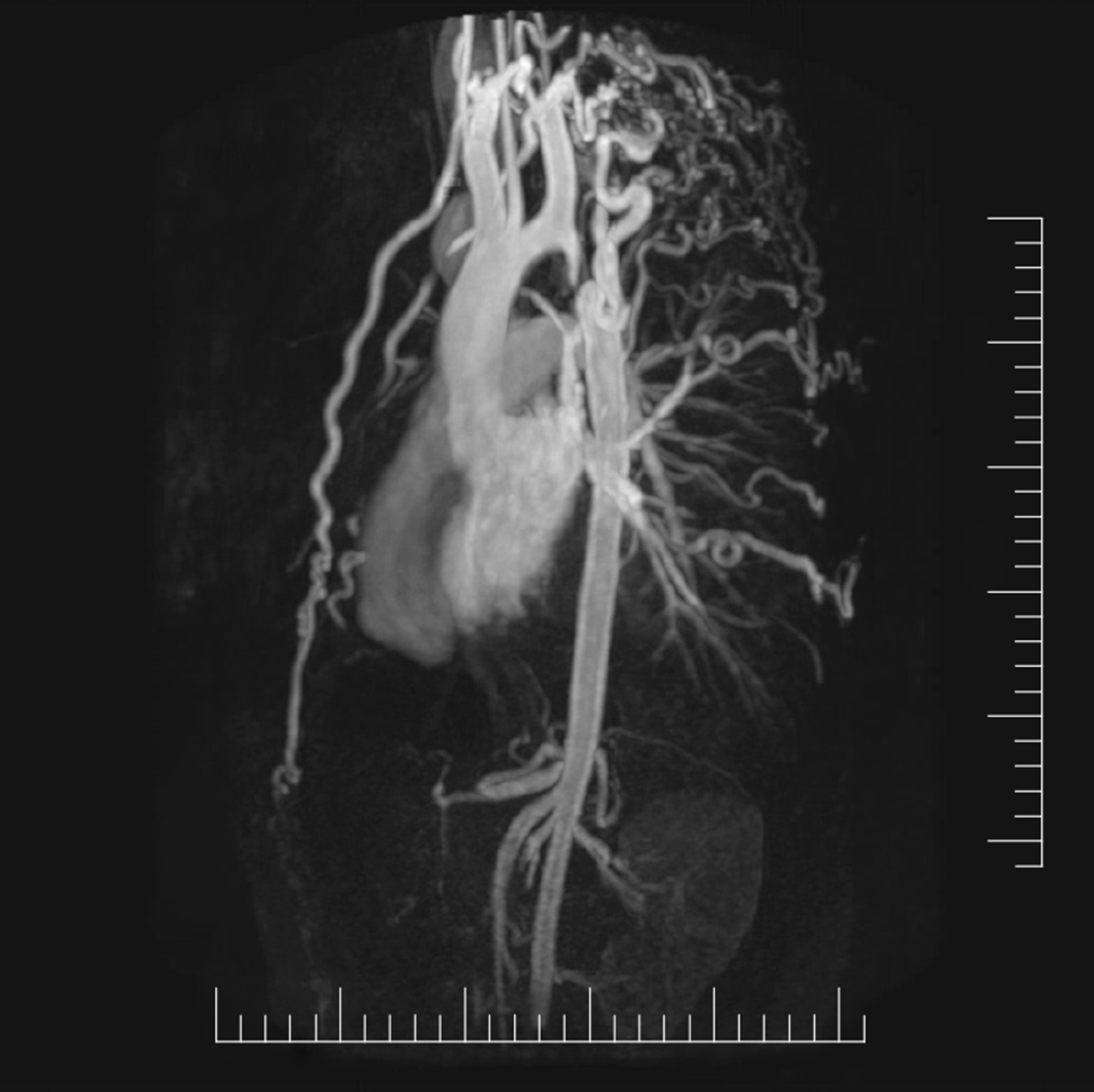
Figure 4 Magnetic resonance imaging demonstrating extensive collaterals in patient with unrepaired coarctation of aorta.
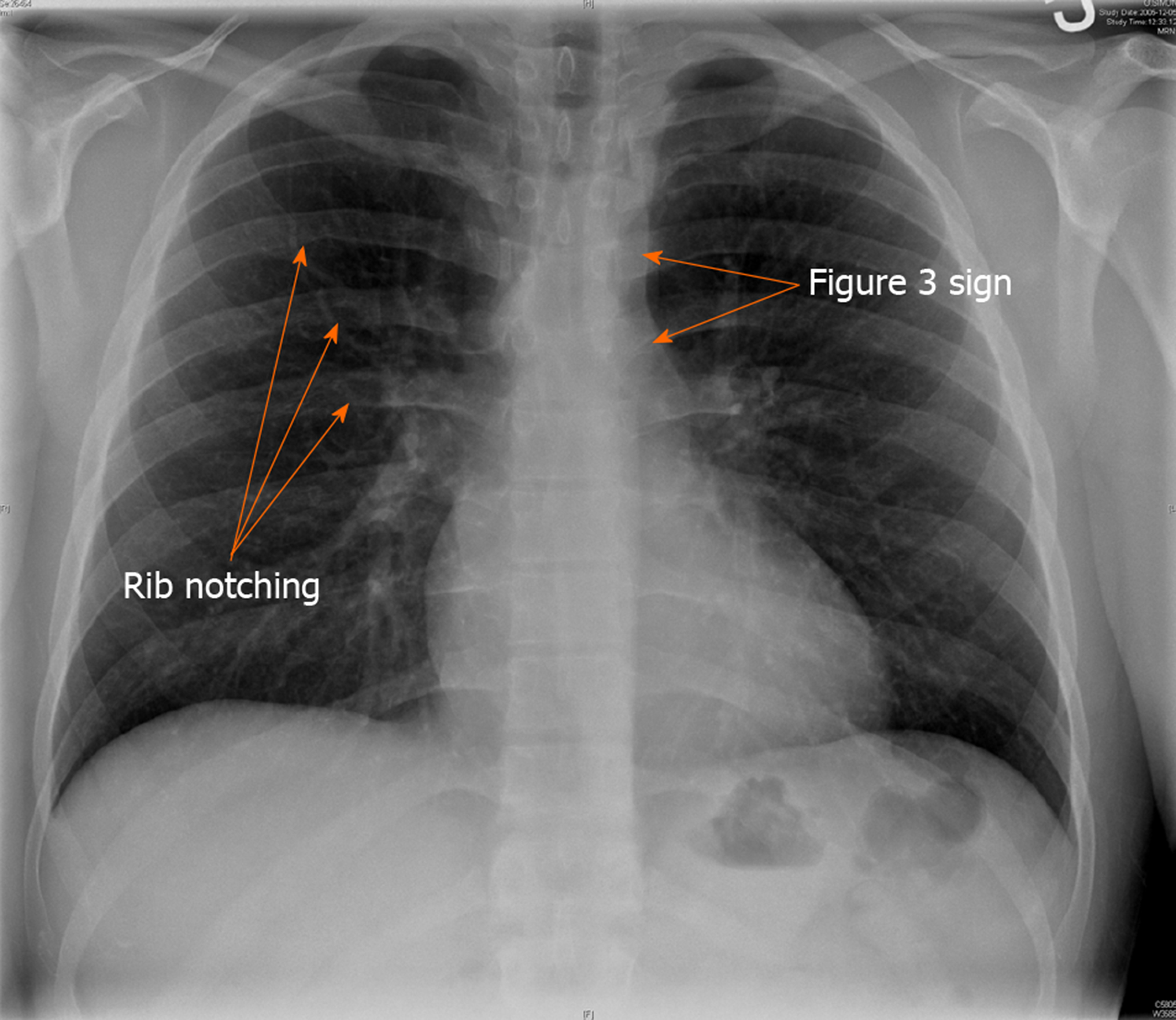
Figure 5 Chest X-Ray demonstrating rib notching and Figure 3 sign.
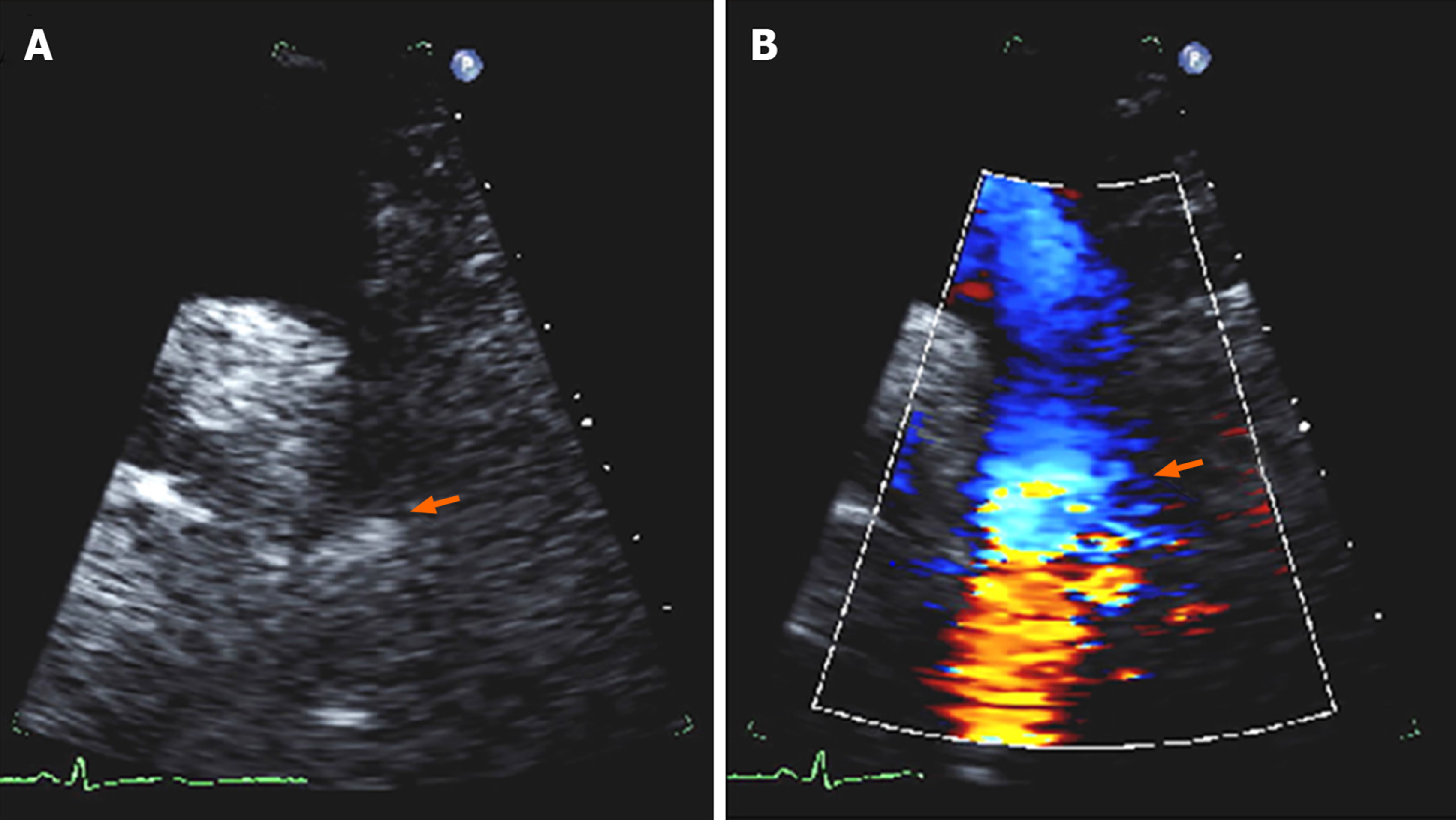
Figure 6 Echocardiographic findings in coarctation of aorta.
A: Two-dimensional suprasternal view on transthoracic echocardiogram demonstrating narrowing in the aortic lumen at the isthmus (arrow); B: Color Doppler imaging demonstrating turbulent flow at the site of the coarctation (arrow).
Figure 7 Doppler echocardiographic findings in coarctation of aorta.
A: Continuous wave Doppler across the coarctation segment in suprastenrtal view demonstrating significant increase in flow velocity with a peak velocity of 4 m/s, comparable to a peak gradient of 64 mmHg based on simplified Bernoulli’s equation; B: Abnormal Doppler pattern in abdominal aorta in a patient with severe coarctation demonstrating blunted velocity with delayed systolic upstroke and continuous diatonic run-off.
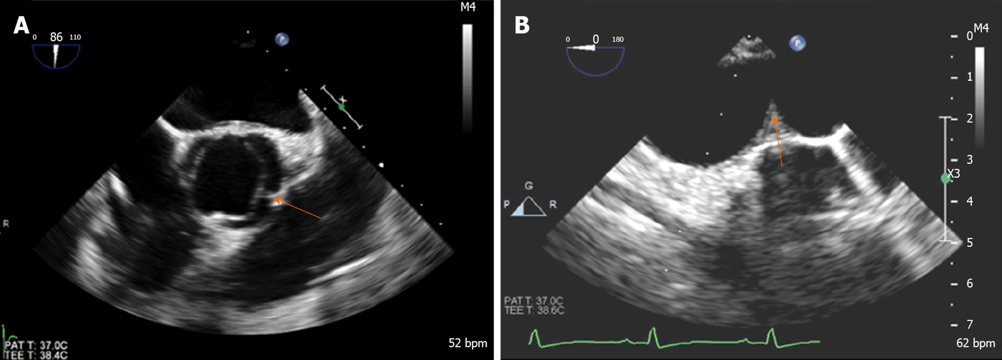
Figure 8 Transesophageal echocardiogram in a patient with coarctation demonstrating.
A: Bicuspid aortic valve with raphe between left and right coronary cusps (arrow); B: Coarctation in distal aortic arch (arrow).
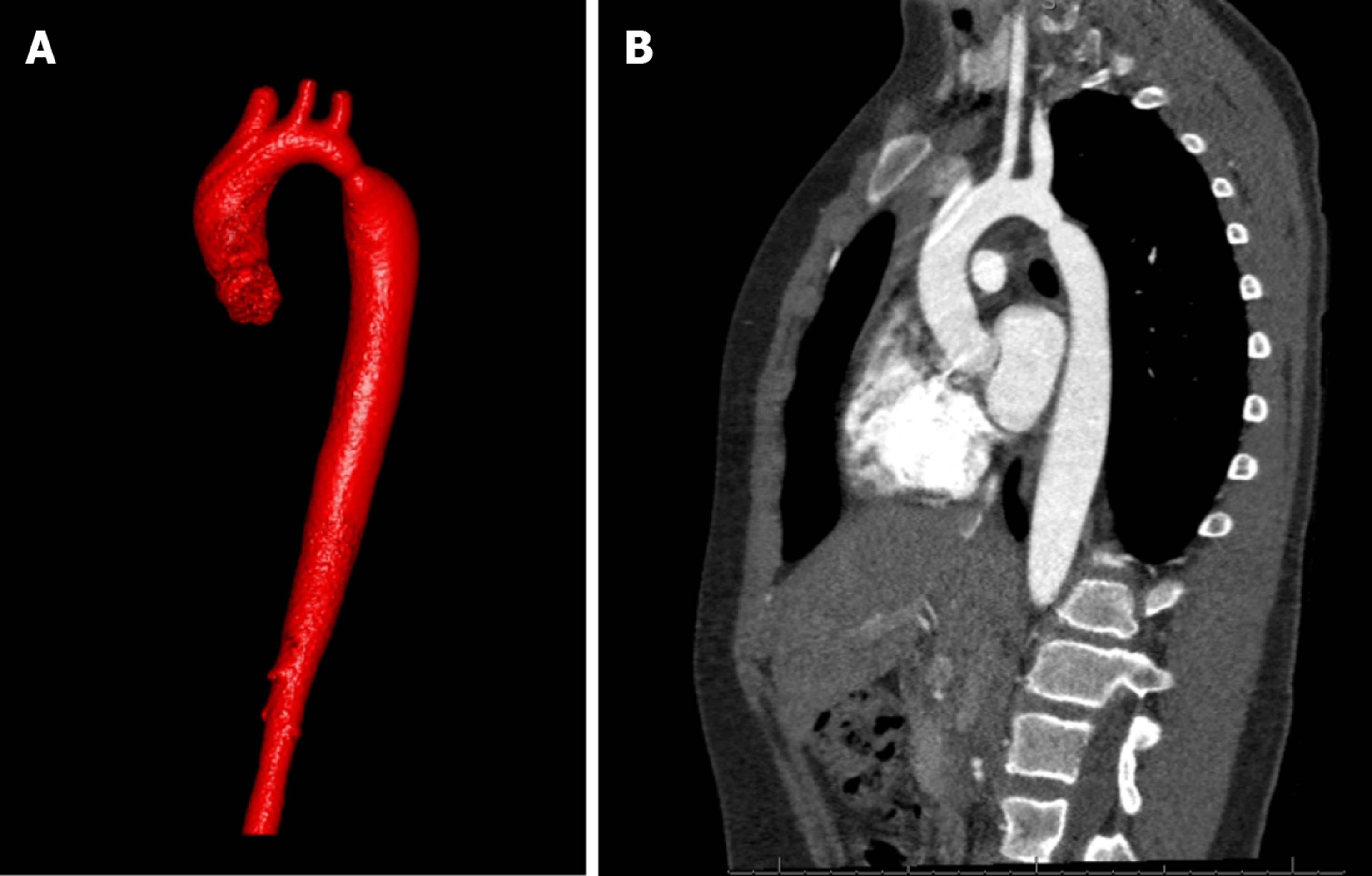
Figure 9 Contrast-enhanced computed tomography demonstrating coarctation of aorta.
A: 3D reconstruction image; B: Sagittal cross-sectional view.
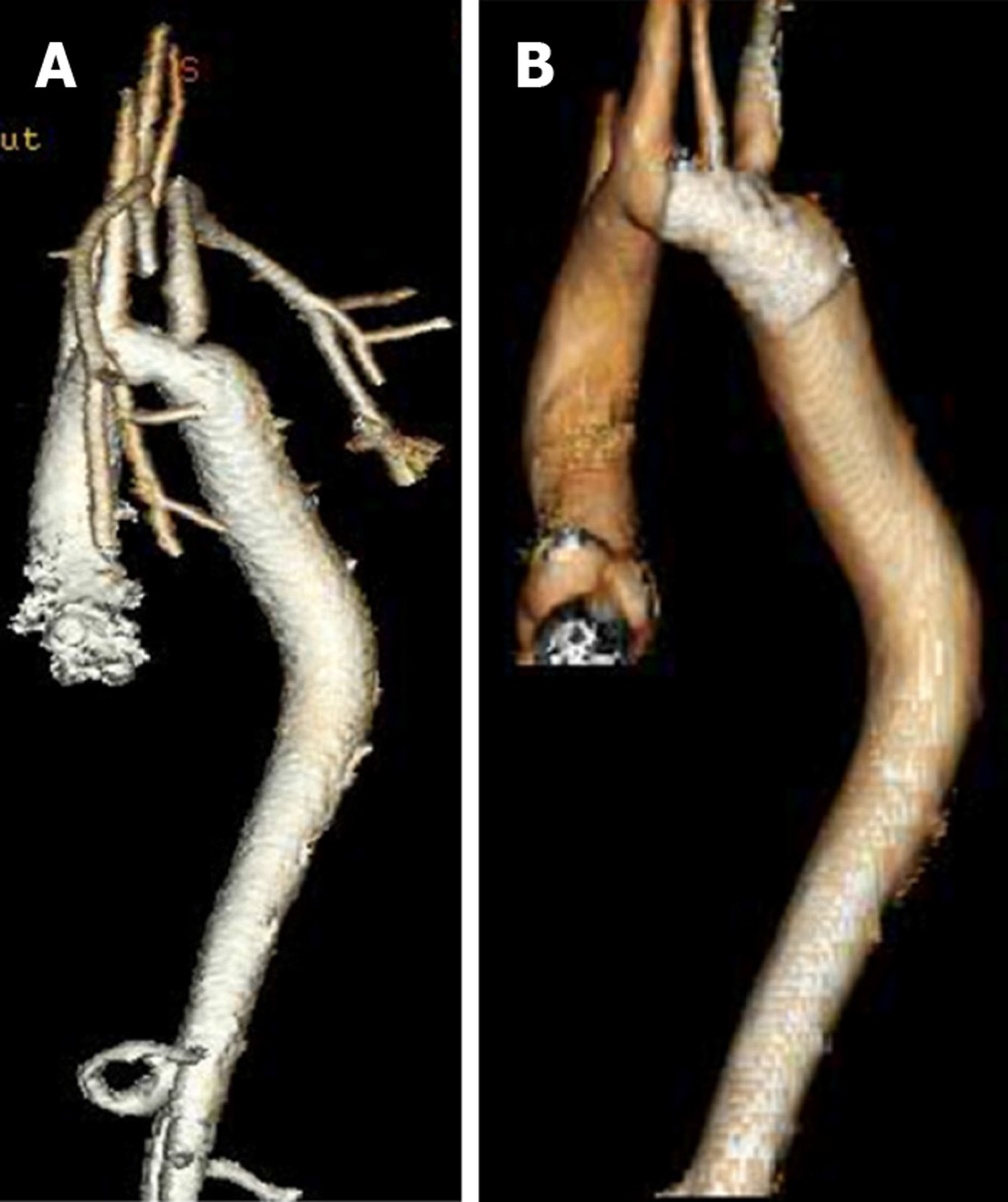
Figure 10 Computed tomography 3D reconstruction images of aortic arch.
A: Pre-intervention; B: Post-intervention.
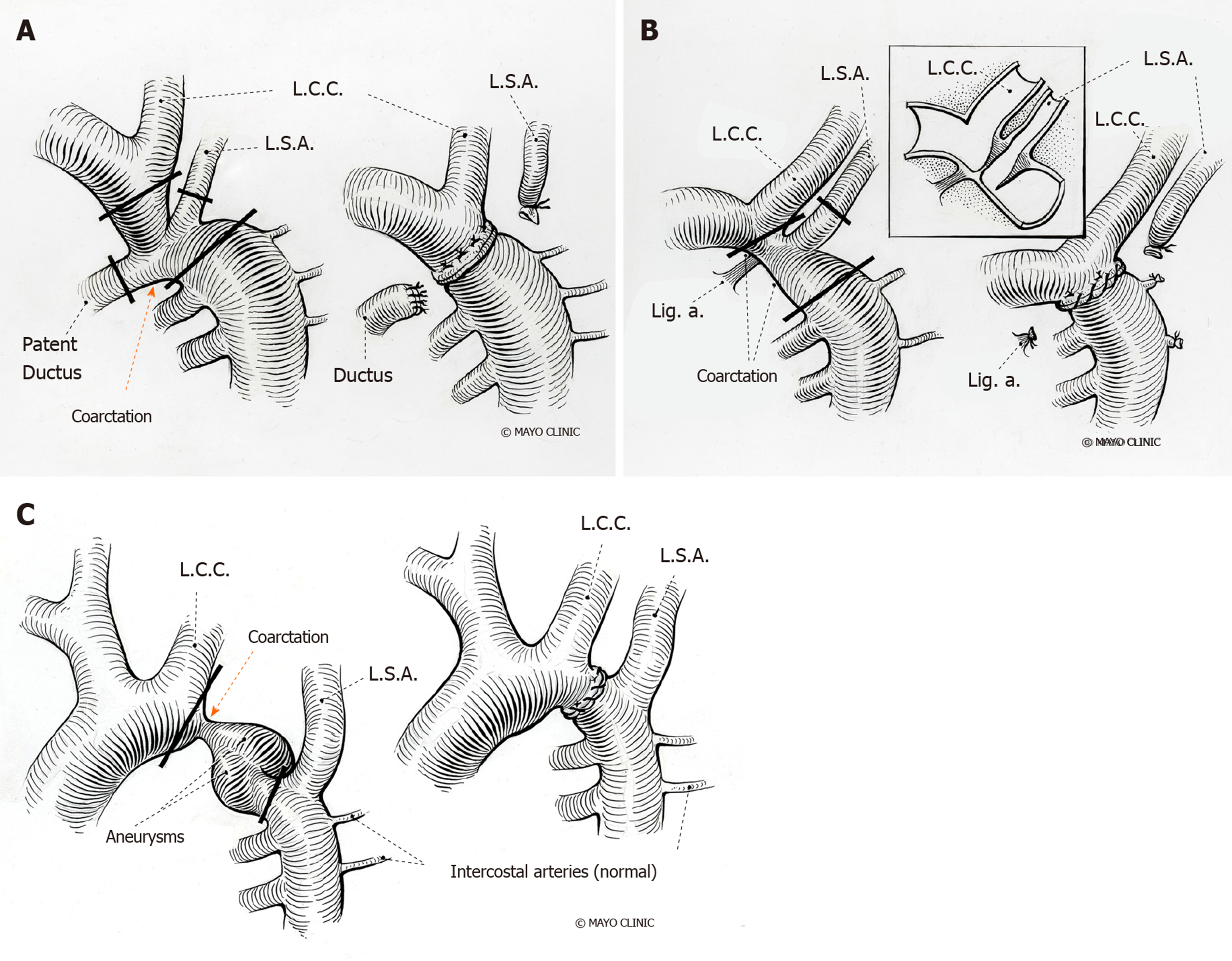
Figure 11 Surgical repair of coarctation of aorta.
A: End to end anastomosis- Resection of the coarctation segment followed by direct suture anastomosis of the transected ends with associated patent ductus arteriosus; B: End-end anastomosis of coarctation without associated patent ductus arteriosus; C: End-end anastomosis of coarctation with associated aneurysm. L.C.C: Left common carotid; L.S.A: Left subclavian artery; Lig. a: Ligamentum arteriosum.
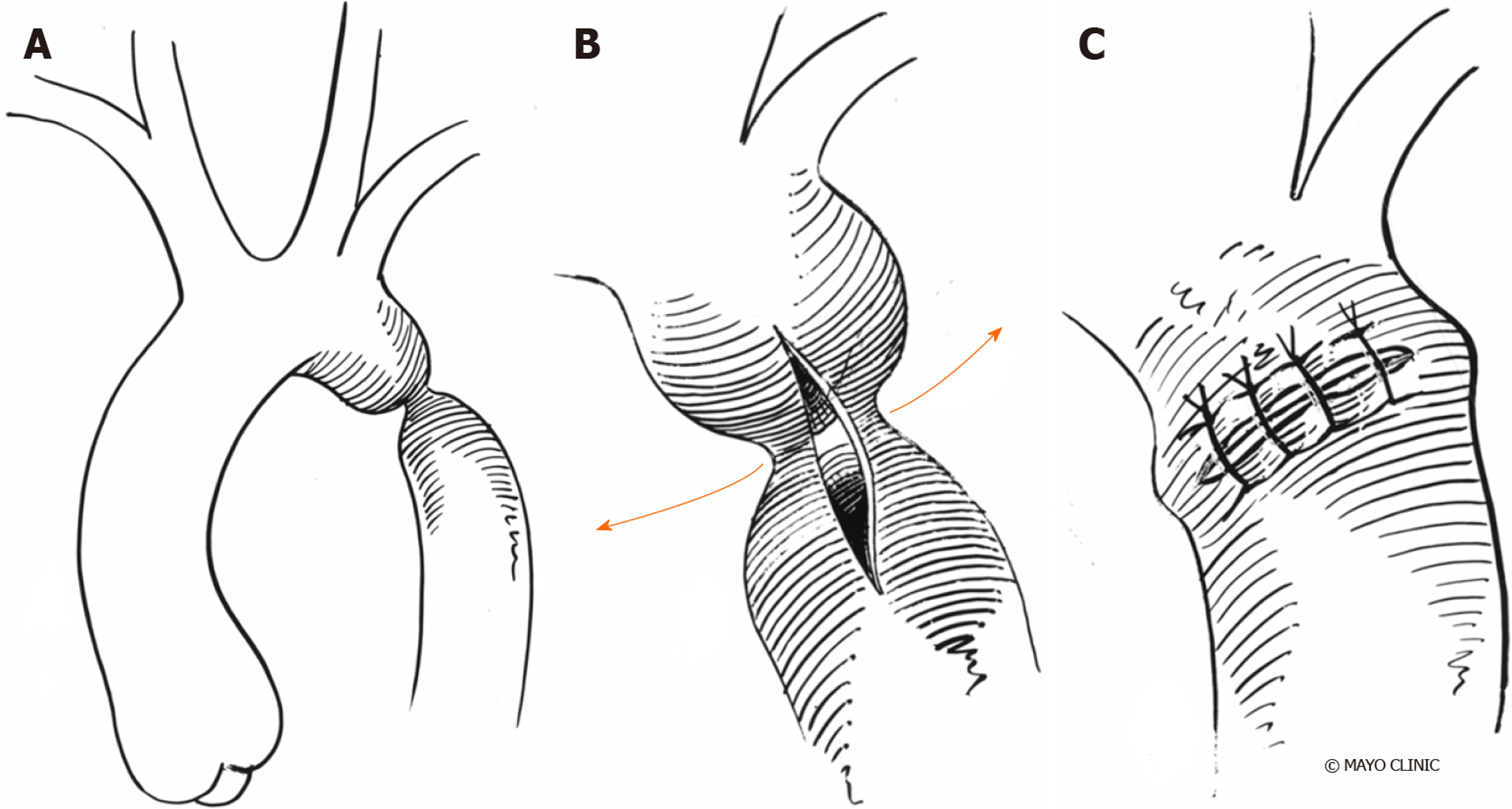
Figure 12 Prosthetic patch aortoplasty.
A: Coarctation before intervention; B: Aorta is incised longitudinally through the coarctation to increase vessel diameter; C: A prosthetic patch used for augmentation of aorta.
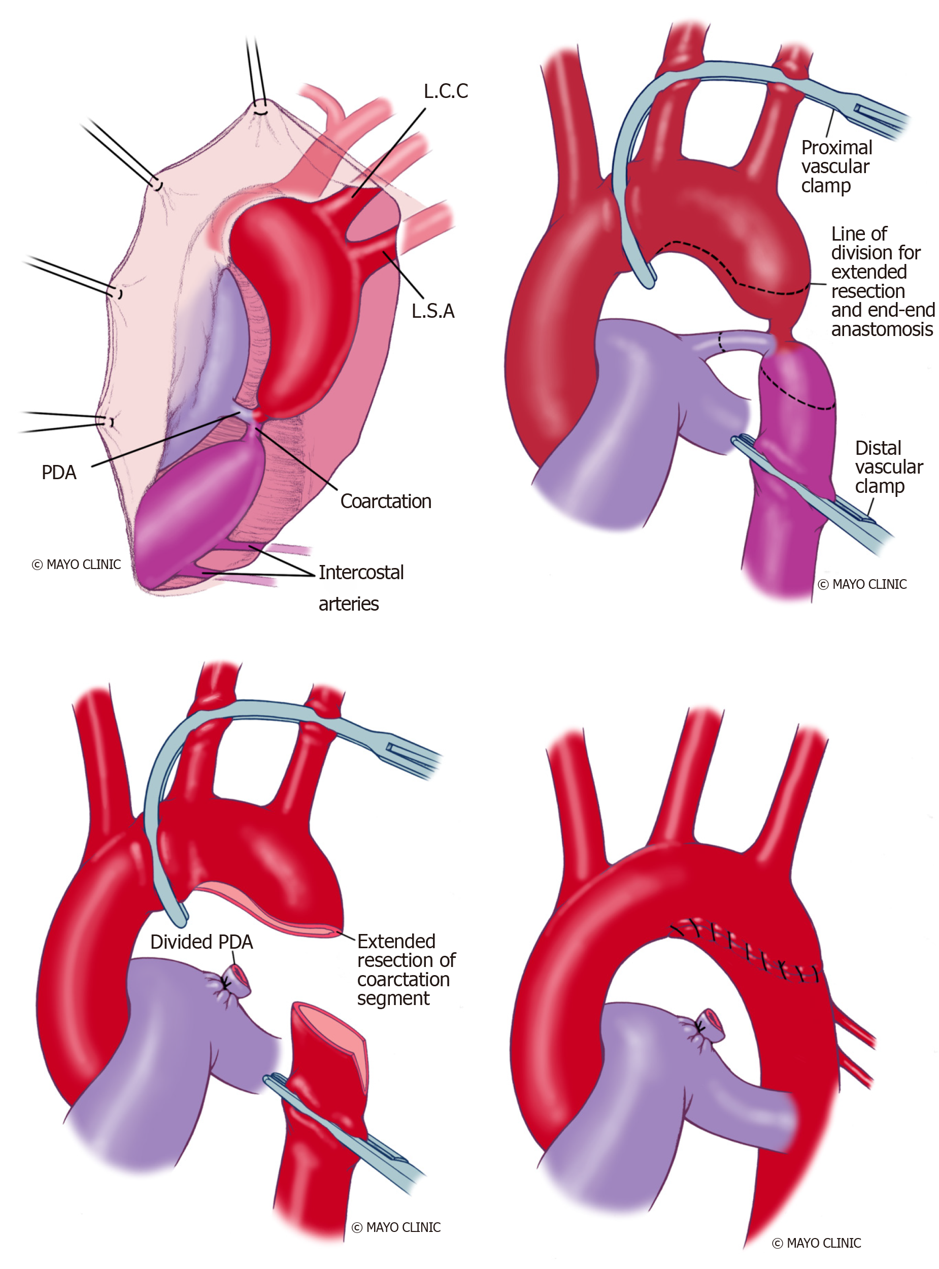
Figure 13 Extended resection and end-end anastomosis.
PDA: Patent ductus Arteriosus; L.C.C: Left common carotid artery; L.S.A: Left subclavian artery.
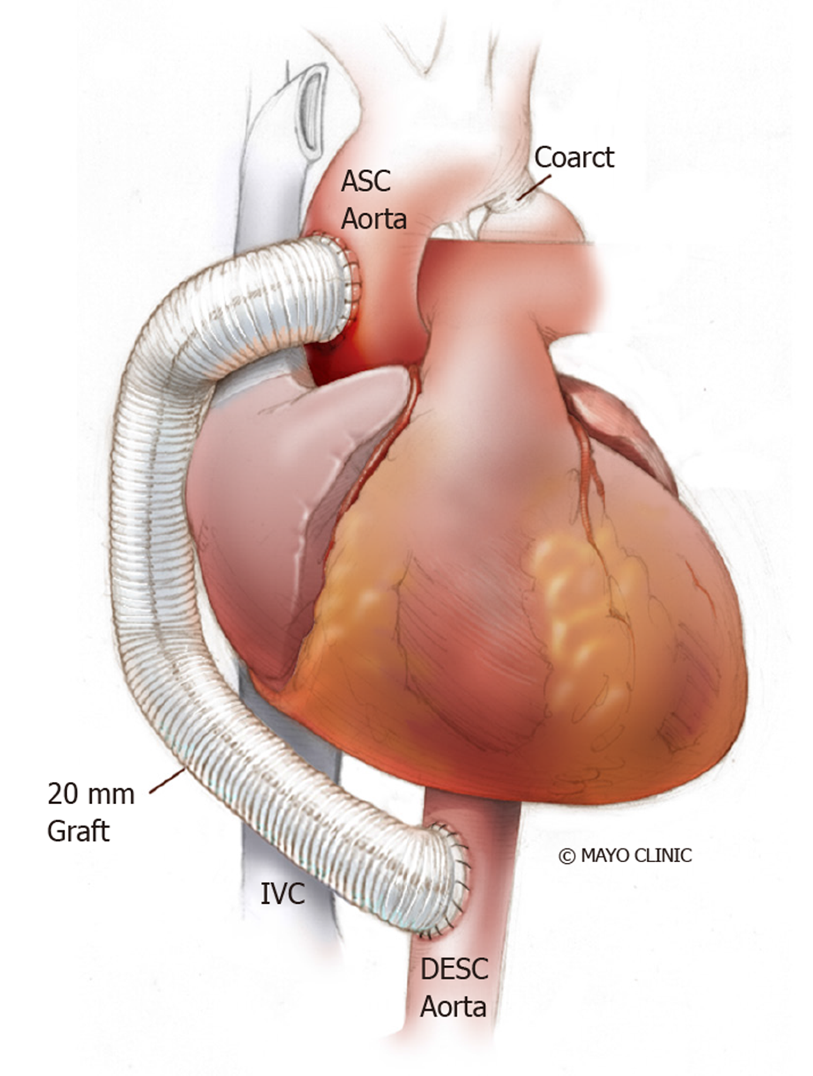
Figure 14 Ascending-to-descending aortic bypass.
ASC: Ascending; IVC: Inferior venacava; DESC: Descending.
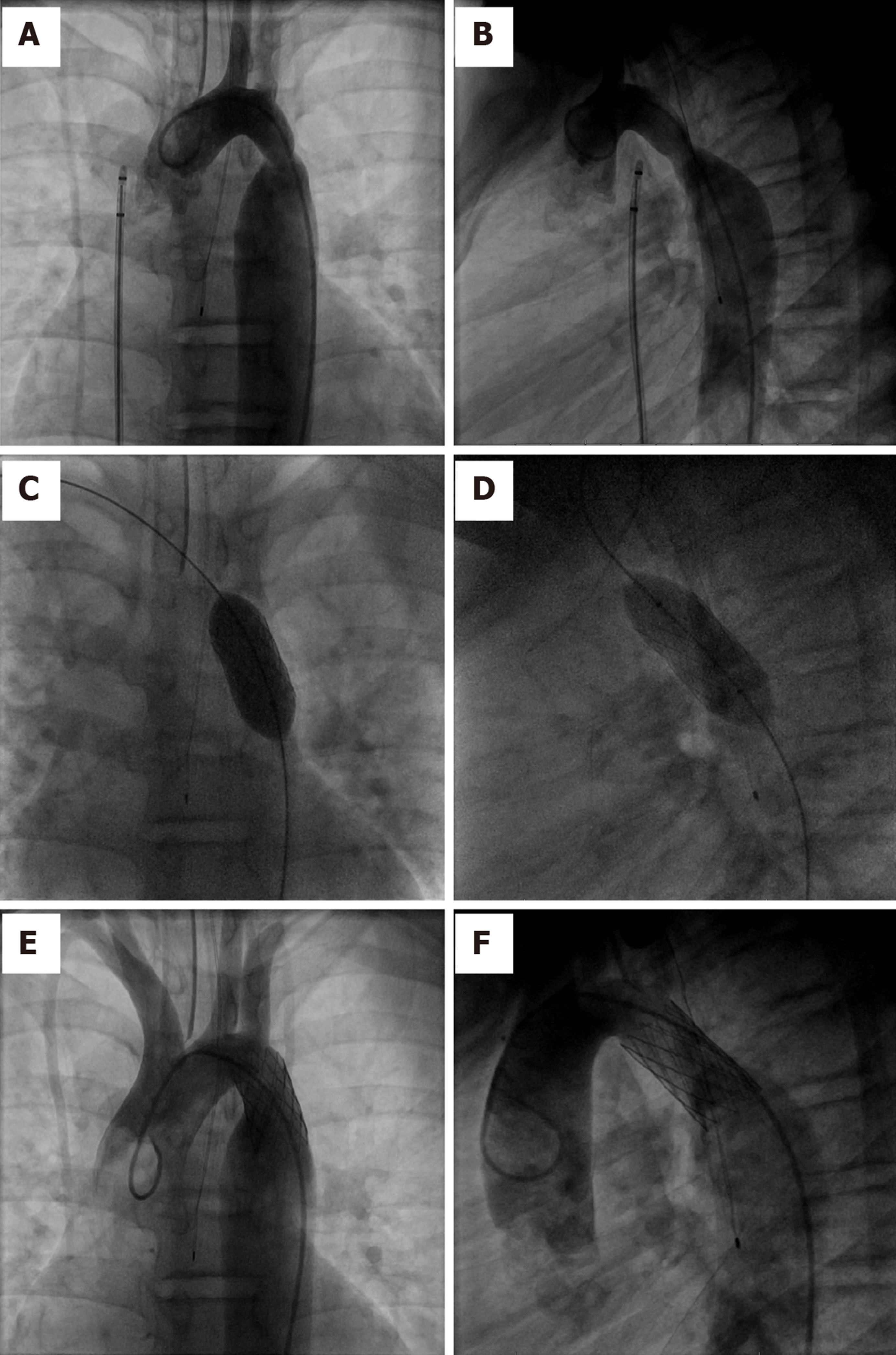
Figure 15 Transcatheter repair of Coarctation of Aorta.
A, B: Angiogram of aortic arch demonstrating coarctation of aorta in antero-posterior and lateral views; C, D: Deployment of covered stent in antero-posterior and lateral views; E, F: Post-intervention aortic arch angiogram demonstrating resolution of coarctation in antero-posterior and lateral views.
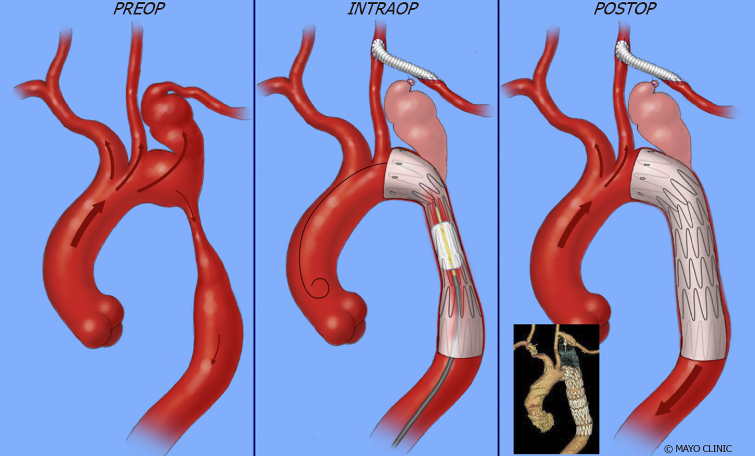
Figure 16 Aneurysm at the origin of left subclavian corrected by deploying a covered stent across the origin of subclavian artery and concomitant surgical anastomosis of left subclavian to left common carotid artery.
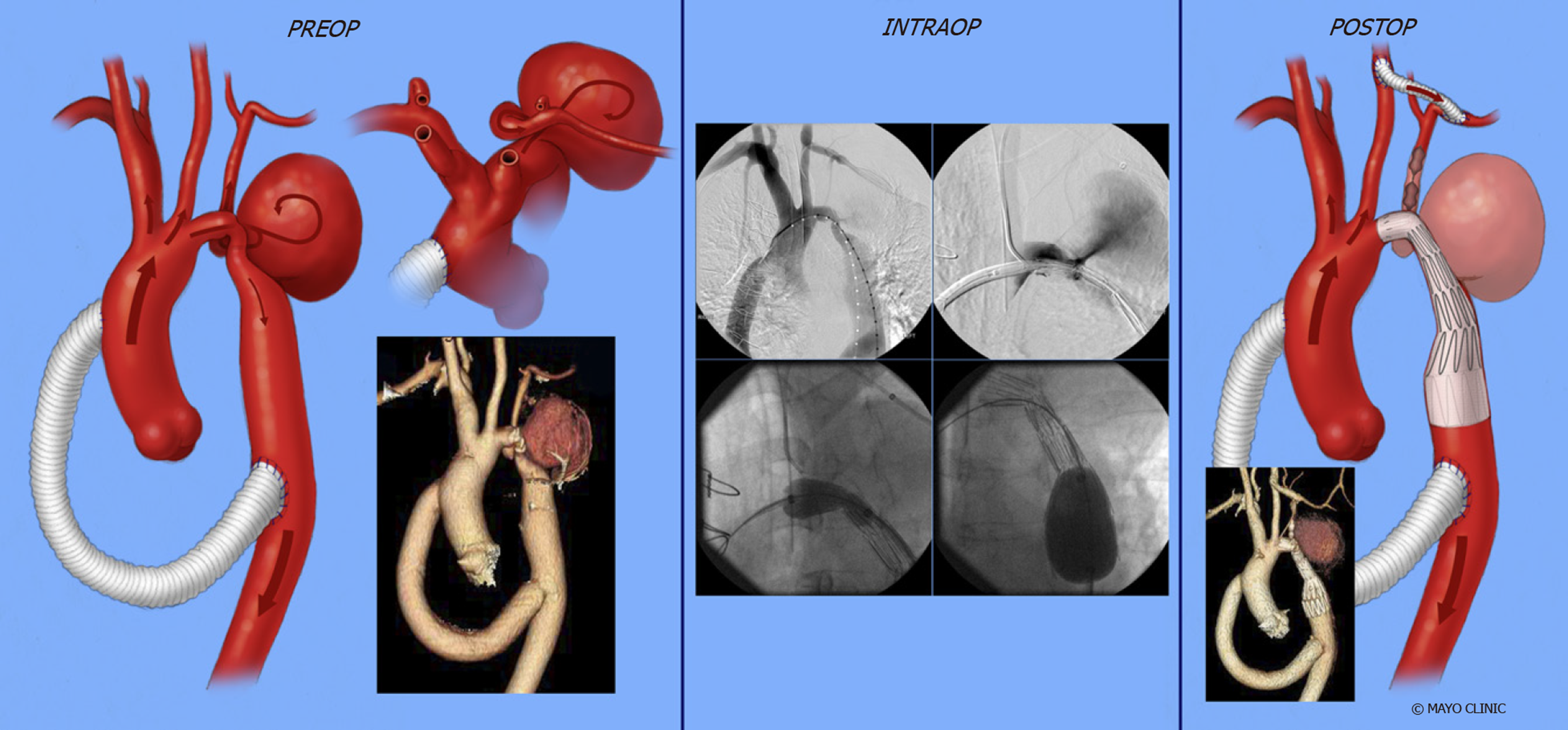
Figure 17 Exclusion of aneurysm of proximal descending thoracic aorta by deploying a covered stent across the aneurysm and concomitant surgical anastomosis of left subclavian to left common carotid artery.
- Citation: Agasthi P, Pujari SH, Tseng A, Graziano JN, Marcotte F, Majdalany D, Mookadam F, Hagler DJ, Arsanjani R. Management of adults with coarctation of aorta. World J Cardiol 2020; 12(5): 167-191
- URL: https://www.wjgnet.com/1949-8462/full/v12/i5/167.htm
- DOI: https://dx.doi.org/10.4330/wjc.v12.i5.167


