Copyright
©The Author(s) 2020.
World J Cardiol. Dec 26, 2020; 12(12): 634-641
Published online Dec 26, 2020. doi: 10.4330/wjc.v12.i12.634
Published online Dec 26, 2020. doi: 10.4330/wjc.v12.i12.634
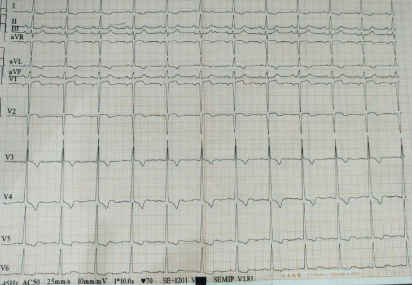
Figure 1 Bedside electrocardiogram after admission.
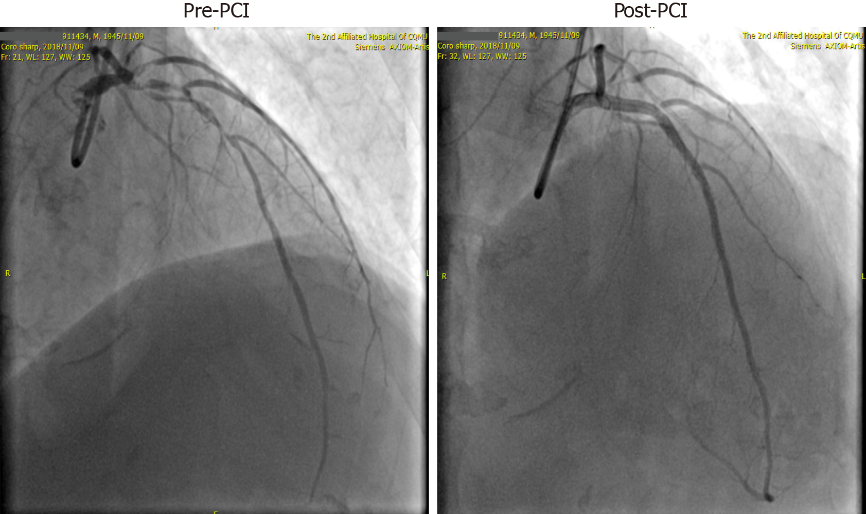
Figure 2 Angiogram showing pre- and post-percutaneous coronary intervention of the left main coronary artery-LAD and left circumflex.
PCI: Percutaneous coronary intervention.
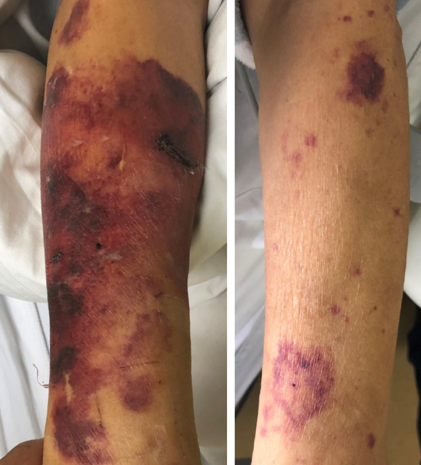
Figure 3 Photograph showing skin necrosis of the right and left upper extremities.
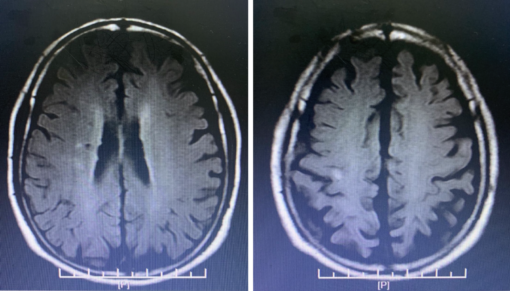
Figure 4 Magnetic resonance imaging showing cerebral infarction.
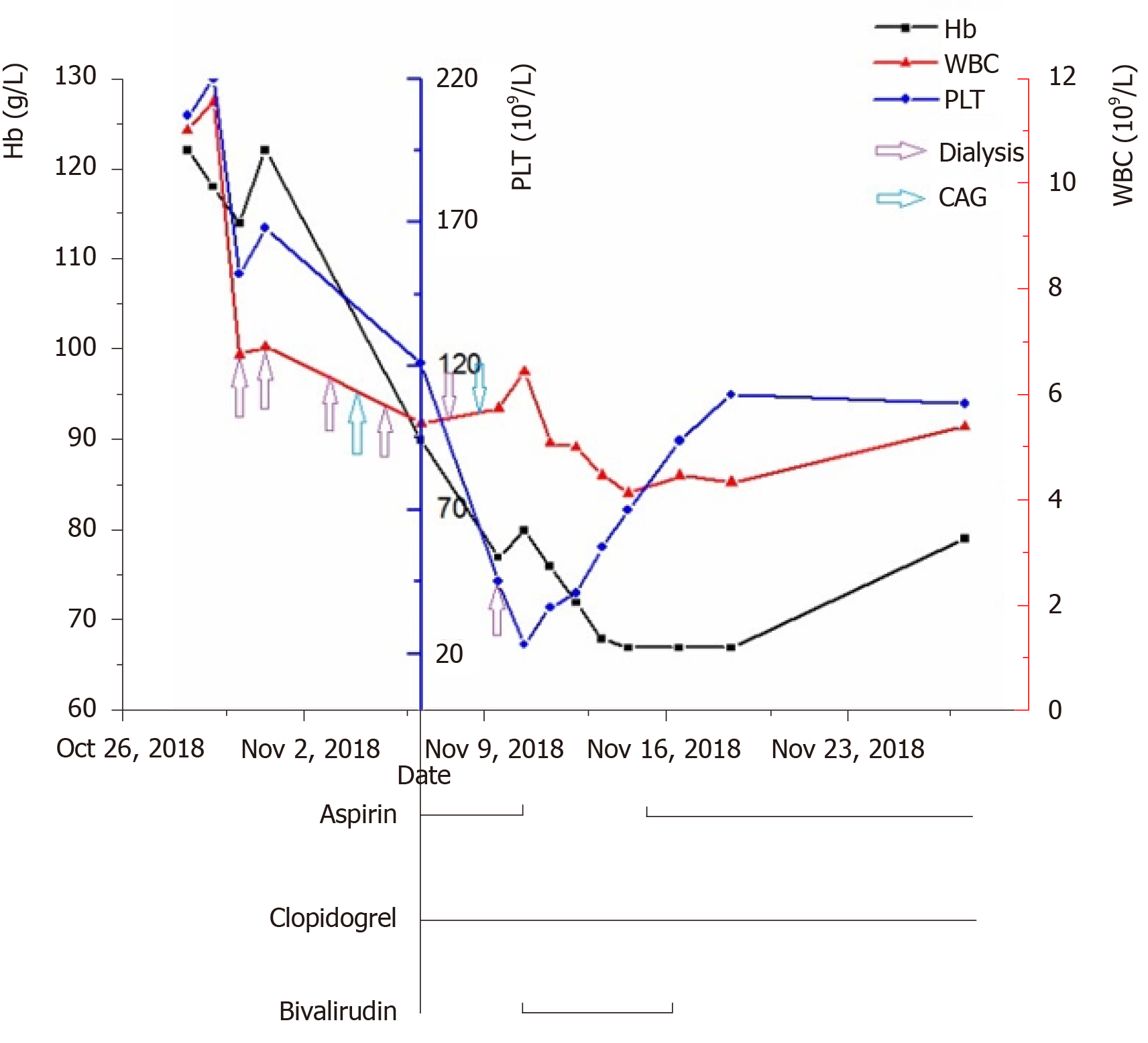
Figure 5 Trends in hemoglobin, white blood cells, and platelets during the patient’s disease course.
Hb: Hemoglobin; WBC: White blood cell; PLT: Platelet; CAG: Coronary angiogram.
- Citation: Wang J, Deng SB, She Q. Heparin-induced thrombocytopenia in renal insufficiency undergoing dialysis and percutaneous coronary intervention after acute myocardial infarction: A case report. World J Cardiol 2020; 12(12): 634-641
- URL: https://www.wjgnet.com/1949-8462/full/v12/i12/634.htm
- DOI: https://dx.doi.org/10.4330/wjc.v12.i12.634


