Copyright
©The Author(s) 2020.
World J Cardiol. Nov 26, 2020; 12(11): 526-539
Published online Nov 26, 2020. doi: 10.4330/wjc.v12.i11.526
Published online Nov 26, 2020. doi: 10.4330/wjc.v12.i11.526
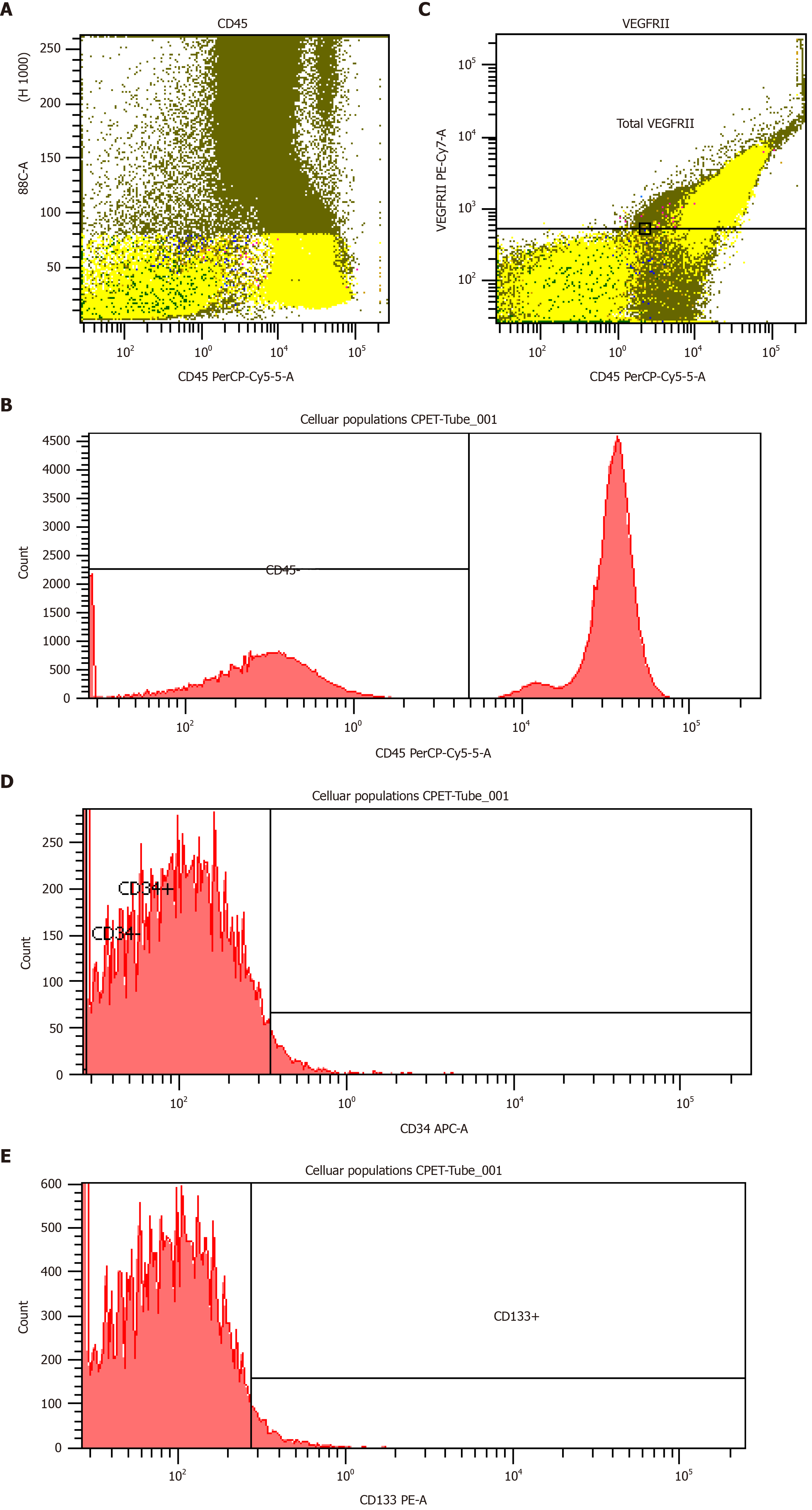
Figure 1 Boolean analysis.
A and B: Flow cytometry analysis for the identification of cellular populations using monoclonal antibodies CD45; C: VEGFR2; D: CD34; E: CD133. In all samples, the CD34 expression was weak. CPET: Cardiopulmonary exercise testing; VEGFR2: Vascular endothelial growth factor receptor 2.
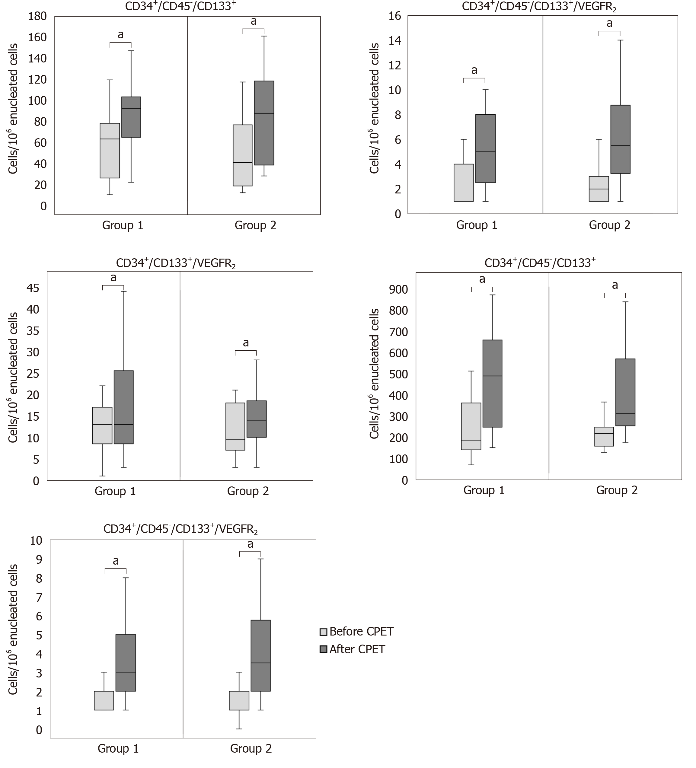
Figure 2 Boxplots representing the acute mobilization of each endothelial cellular population before and after a symptom limited maximal cardiopulmonary exercise testing between two severity groups according to the median value of peak oxygen uptake.
Group 1: Peak oxygen uptake (VO2) < 18.0 mL/kg/min; Group 2: Peak VO2 ≥ 18.0 mL/kg/min). aP < 0.05 indicates statistically significantly increase.
- Citation: Kourek C, Karatzanos E, Psarra K, Georgiopoulos G, Delis D, Linardatou V, Gavrielatos G, Papadopoulos C, Nanas S, Dimopoulos S. Endothelial progenitor cells mobilization after maximal exercise according to heart failure severity. World J Cardiol 2020; 12(11): 526-539
- URL: https://www.wjgnet.com/1949-8462/full/v12/i11/526.htm
- DOI: https://dx.doi.org/10.4330/wjc.v12.i11.526


