Copyright
©The Author(s) 2019.
World J Cardiol. Jul 26, 2019; 11(7): 189-194
Published online Jul 26, 2019. doi: 10.4330/wjc.v11.i7.189
Published online Jul 26, 2019. doi: 10.4330/wjc.v11.i7.189
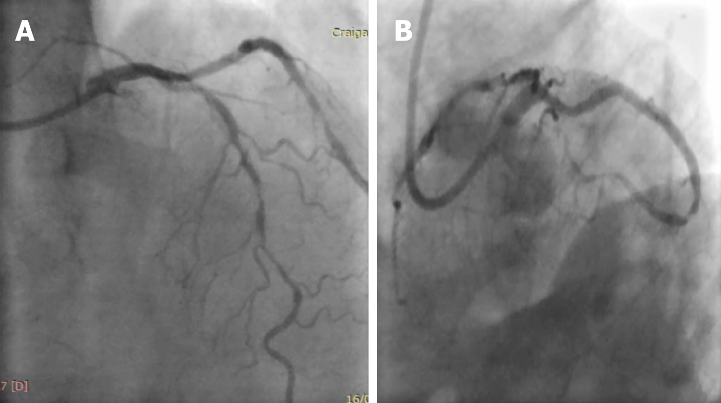
Figure 1 Left coronary angiography demonstrating heavily calcified left anterior descending artery.
A: Cranial projection; B: Caudal projection. Excellent results from previous percutaneous coronary intervention to LCx.
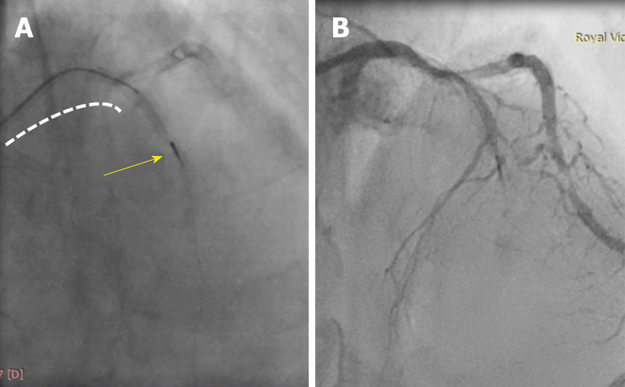
Figure 2 Fractured micro-catheter tip.
A: The yellow arrow points at the trapped corsair tip in mid left anterior descending artery (LAD) with the remaining micro-catheter in the proximal LAD segment (dotted white line); B: No flow in the LAD as a result of the jailed tip.
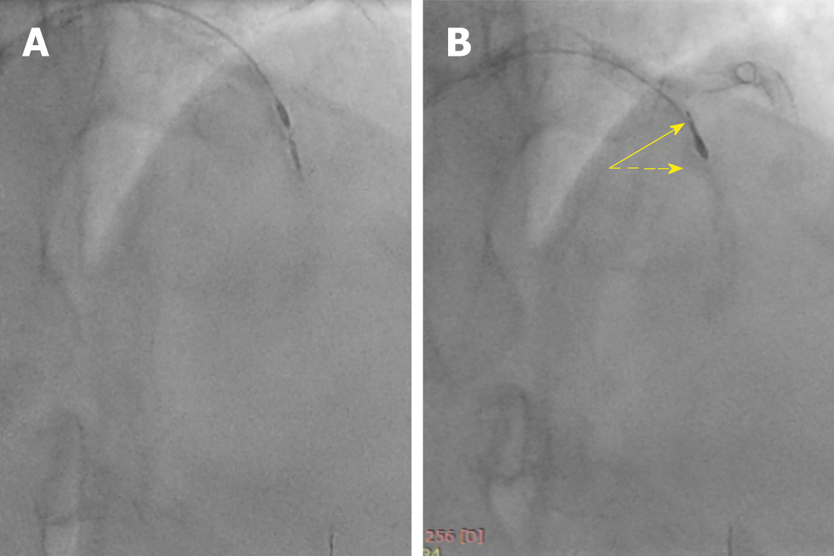
Figure 3 Rotational atherectomy was performed to modify calcification adjacent to the trapped tip.
A: Calcium modifications using rota burr; B: Corsair tip was freed up and directed towards small diagonal branch.
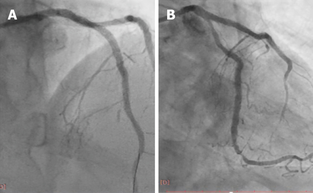
Figure 4 Final angiographic results.
A: Good flow in the left anterior descending artery with occluded diagonal branch by the corsair tip in the cranial projection; B: Caudal projection demonstrating final angiographic results.
- Citation: Alkhalil M, McQuillan C, Moore M, Spence MS, Owens C. Use of rotablation to rescue a “fractured” micro catheter tip: A case report. World J Cardiol 2019; 11(7): 189-194
- URL: https://www.wjgnet.com/1949-8462/full/v11/i7/189.htm
- DOI: https://dx.doi.org/10.4330/wjc.v11.i7.189


