Copyright
©The Author(s) 2019.
World J Cardiol. Oct 26, 2019; 11(10): 221-235
Published online Oct 26, 2019. doi: 10.4330/wjc.v11.i10.221
Published online Oct 26, 2019. doi: 10.4330/wjc.v11.i10.221
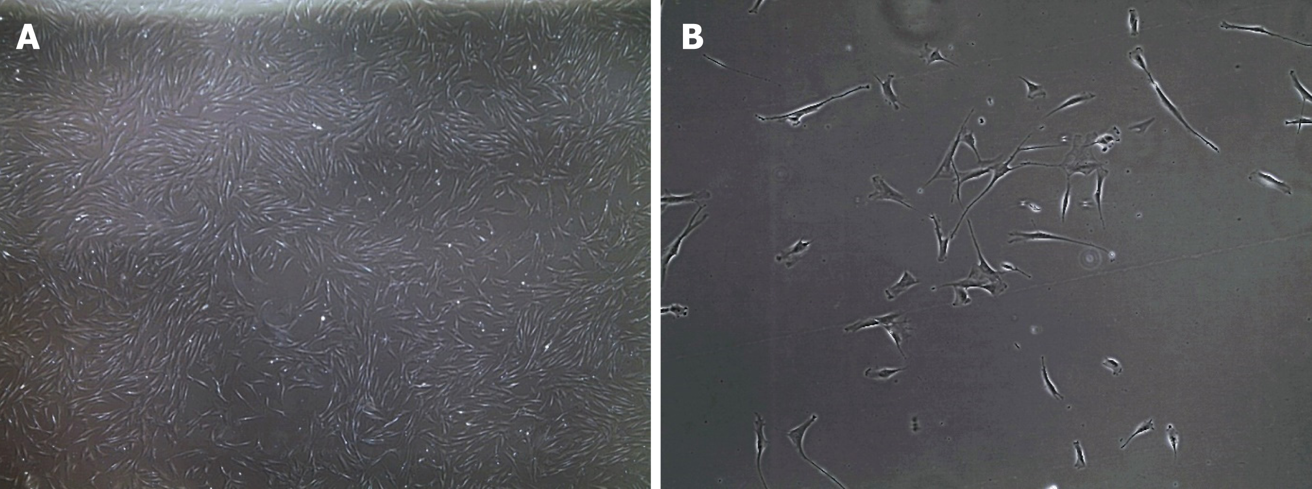
Figure 1 Bright field microscopy images of human fibroblasts.
A: 4 × magnification; and B: 20 × magnification.
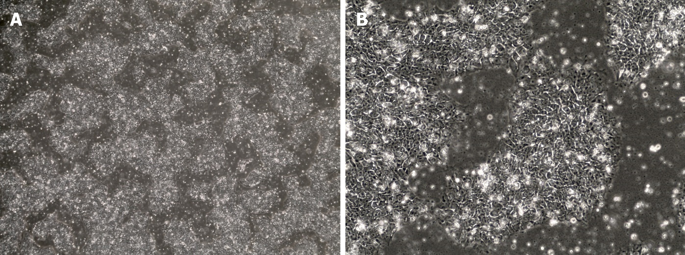
Figure 2 Bright field microscopy images of human induced pluripotent stem cells.
Cells display a round morphology with a large nucleus and grow firmly packed in colonies. A: 4 × magnification. B: 20 × magnification.
Figure 3 Cardiomyocyte differentiation protocol.
Modified from Lian et al[135], 2012. hiPSCs: Human induced pluripotent stem cells.
- Citation: Jimenez-Tellez N, Greenway SC. Cellular models for human cardiomyopathy: What is the best option? World J Cardiol 2019; 11(10): 221-235
- URL: https://www.wjgnet.com/1949-8462/full/v11/i10/221.htm
- DOI: https://dx.doi.org/10.4330/wjc.v11.i10.221


