Copyright
©The Author(s) 2018.
World J Cardiol. Oct 26, 2018; 10(10): 187-190
Published online Oct 26, 2018. doi: 10.4330/wjc.v10.i10.187
Published online Oct 26, 2018. doi: 10.4330/wjc.v10.i10.187
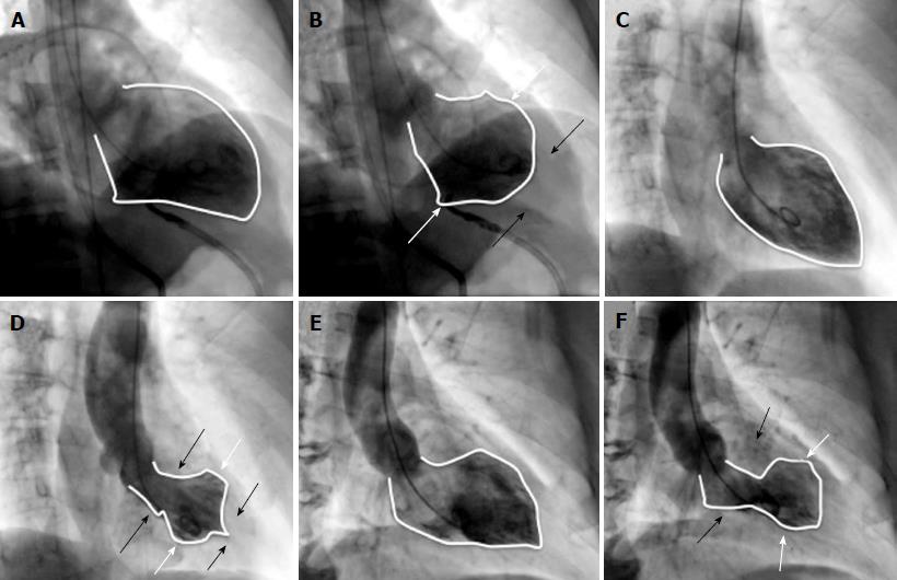
Figure 1 Ventriculography of the 3 cases.
A and B are the ventriculography of case 1. A: LV at end diastole; B: LV at end systole with motion defect (white arrows show inferior and anterior basal hypokinesis; black arrows show hypercontractility of apical segments); LVEF: 20%. C and D are the ventriculography of case 2. C: LV at end diastole; D: LV at end systole with motion defect (white arrows show inferior and anterior midventricular hypokinesis; black arrows show hypercontractility of inferior and anterior basal and apical segments); LVEF: 60%. E and F are the ventriculography of case 3; E: LV at end diastole; F: LV at end systole with apical ballooning (white arrows show apical inferior and anterior hypokinesis; black arrows show inferior and anterior basal segments hypercontractility); LVEF: 45%, with severe mitral regurgitation. LV: Left ventricle; LVEF: Left ventricular ejection fraction.
- Citation: Fuensalida A, Cortés M, Gabrielli L, Méndez M, Martínez A, Martínez G. Takotsubo syndrome - different presentations for a single disease: A case report and review of literature. World J Cardiol 2018; 10(10): 187-190
- URL: https://www.wjgnet.com/1949-8462/full/v10/i10/187.htm
- DOI: https://dx.doi.org/10.4330/wjc.v10.i10.187


