Copyright
©The Author(s) 2018.
World J Cardiol. Oct 26, 2018; 10(10): 145-152
Published online Oct 26, 2018. doi: 10.4330/wjc.v10.i10.145
Published online Oct 26, 2018. doi: 10.4330/wjc.v10.i10.145
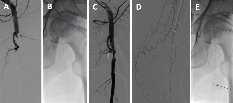
Figure 1 The angiography of patient A.
A: Thrombotic occlusion of the common femoral artery (CFA) (blue arrow); B: Mechanical debulking using the Rotarex®S catheter (arrow); C,D: Anterograde flow was restored in the CFA after debulking with reduced flow in the foot arteries; E: Local thrombolysis was performed using a dedicated Unifuse catheter (arrow).
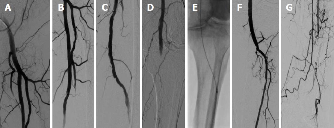
Figure 2 The angiography of patient A.
The following day, a complete resolution of thrombus material was found in the common femoral artery. A-D: Superficial femoral artery and deep femoral artery (A-C) with new occlusion of the popliteal artery (D); E: Repeated Rotarex® debulking in the popliteal artery and continued to the proximal and median parts of the anterior tibial artery; F,G: Restoration of flow of the anterior tibial artery and of the foot.
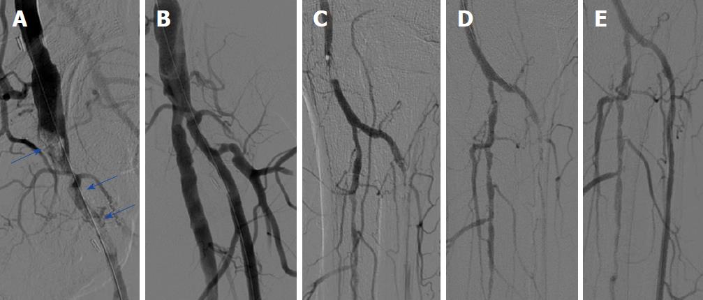
Figure 3 The angiography of patient B.
A: Thrombotic occlusion of the common femoral artery and profound femoral artery (blue arrows); B: Good angiographic reflow after repeated treatment with the 6F Rotarex®S mechanical debulking catheter; C: Thrombus formation in the popliteal and in the anterior tibial artery; D: No flow restoration in the anterior tibial artery after treatment with the 6F Rotarex®S in the popliteal artery; E: Flow restoration after deploying the Rotarex®S in the anterior tibial artery.
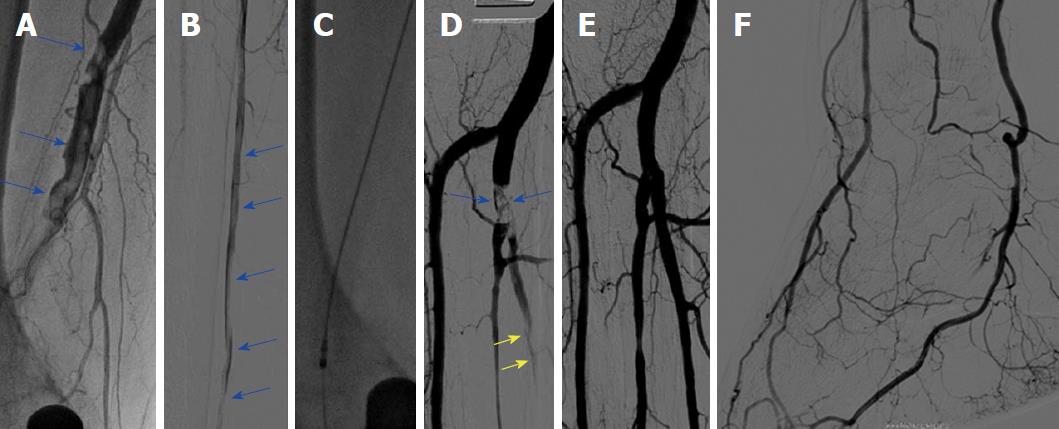
Figure 4 The angiography of patient C.
A: Thrombotic occlusion of the right popliteal artery (blue arrows); B: Further thrombus formation in the right posterior tibial artery (blue arrows); C: Rotarex®S catheter in the popliteal artery; D: Remaining thrombus formation in the tibiofibular trunk and in the posterior tibial artery (arrows); E,F: Flow restoration in the foot after using the Rotarex®S in the proximal and median segments of the posterior tibial artery.
- Citation: Giusca S, Raupp D, Dreyer D, Eisenbach C, Korosoglou G. Successful endovascular treatment in patients with acute thromboembolic ischemia of the lower limb including the crural arteries. World J Cardiol 2018; 10(10): 145-152
- URL: https://www.wjgnet.com/1949-8462/full/v10/i10/145.htm
- DOI: https://dx.doi.org/10.4330/wjc.v10.i10.145


