Copyright
©The Author(s) 2018.
World J Cardiol. Oct 26, 2018; 10(10): 127-140
Published online Oct 26, 2018. doi: 10.4330/wjc.v10.i10.127
Published online Oct 26, 2018. doi: 10.4330/wjc.v10.i10.127
Figure 1 Wrap-around left anterior descending artery.
A, B: Angiographic views of a large left anterior descending artery that wraps around the apex of the heart and supplies blood to part of the inferior wall (arrows) in anteroposterior caudal (A) and cranial view (B). From the authors’ personal dataset.
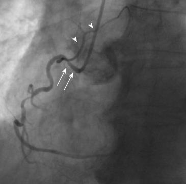
Figure 2 Shepherd’s crook right coronary artery.
Arrows show the prominent ascending course of proximal right coronary artery segment and an equally prominent, almost 180° switchback turn (left anterior oblique view). Arrowheads show the sinoatrial branch. From the authors’ personal dataset.
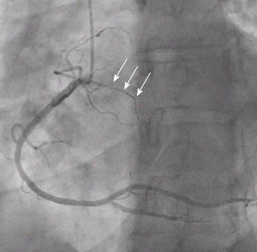
Figure 3 Descending septal branch.
A branch of proximal right coronary artery (arrows) providing blood to part of the basal interventricular septum (left anterior oblique view with shallow cranial angulation). This branch may occasionally have a separate origin from the right sinus of Valsalva. From the authors’ personal dataset.
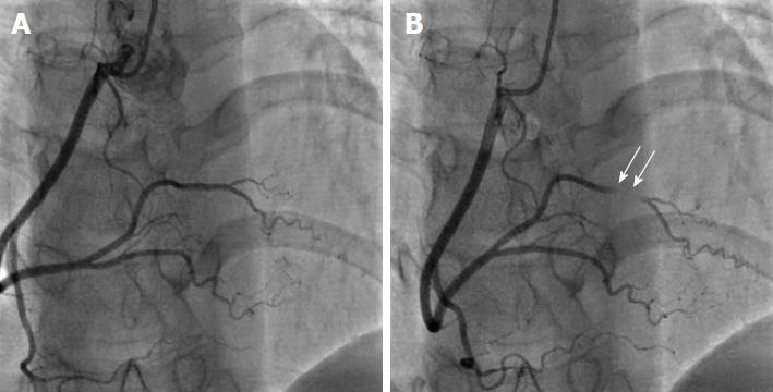
Figure 4 Myocardial bridging.
A: Anteroposterior view with cranial angulation shows normal appearance of right coronary artery in diastole; B: Complete obliteration of a short segment of the posterolateral branch in systole (arrows) due to myocardial bridging. From the authors’ personal dataset.
Figure 5 Absence of left main stem.
A, B: Separate ostia of left circumflex artery (A, anteroposterior caudal view) and left anterior descending artery (B, shallow right anterior oblique view with cranial angulation) indicating a congenital absence of left main stem. From the authors’ personal dataset.
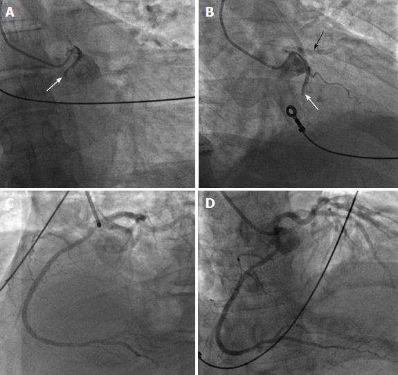
Figure 6 Ectopic right coronary artery originating from the left sinus of Valsalva.
A, B: Proximal occlusion of anomalous right coronary artery (RCA) (white arrows) with origin from the left sinus of Valsalva in a patient with inferior wall ST elevation myocardial infarction (A: anteroposterior caudal view; B: right anterior oblique view). The black arrow in panel B shows a patent proximal left anterior descending artery in non-selective angiography of the left coronary artery (LCA); C, D: Left anterior oblique view with cranial angulation (C) and anteroposterior cranial view (D) show the LCA and an excellent final angiographic of the anomalous RCA after successful primary percutaneous coronary intervention. From the authors’ personal data and from Ref.[73] (modified with permission).
Figure 7 Ectopic left anterior descending artery originating from the right sinus of Valsalva.
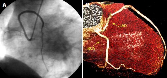
A: Left anterior oblique view with cranial angulation of the left anterior descending artery (LAD) originating from the right Valsalva sinus in coronary angiogram; B: Reconstructed image from computed tomography coronary angiogram showing both LAD and right coronary artery arising from the right sinus of Valsalva. From the authors’ personal dataset. LAD: Left anterior descending; RCA: Right coronary artery.
Figure 8 Ectopic left circumflex artery originating from the right sinus of Valsalva.
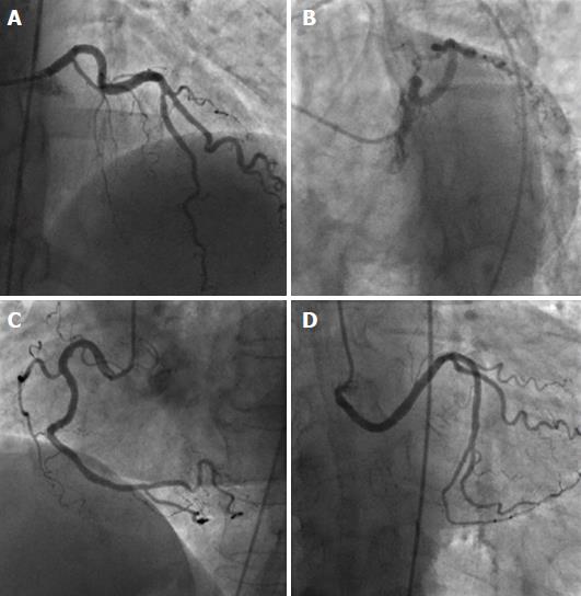
A, B: Left coronary artery providing left anterior descending artery, with absence of left circumflex artery (LCX) (A: anteroposterior cranial view; B: left anterior oblique view with caudal angulation); C, D: Separate origin of right coronary artery (C, left anterior oblique view) and aberrant LCX from the right sinus of Valsava (D, anteroposterior caudal view). From the authors’ personal dataset.
Figure 9 Anomalous origin of the left coronary artery from the pulmonary artery in infant.
A: Cardiomegaly in chest X-ray; B: Aortogram showing only right coronary artery (RCA) originating from the aortic sinuses (arrowhead) but no left coronary artery; C: Selective angiography of RCA shows extensive collaterals from the RCA backfilling the left system and the pulmonary artery (arrows). Reproduced from ref. [74].
Figure 10 Anomalous origin of the left coronary artery from the pulmonary artery in adult.
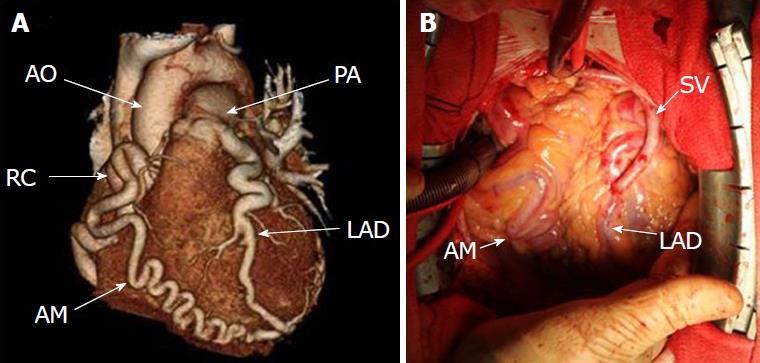
A: Three-dimensional reconstruction of computed tomography images showing prominent collaterals from right coronary artery to the left anterior descending artery (LAD), while LAD originates from the pulmonary artery; B: Coronary bypass grafting with a saphenous vein graft to the LAD. Reproduced from ref. [75]. SV: Saphenous vein; RC: Right coronary; LAD: Left anterior descending.
Figure 11 Single coronary artery from the left sinus.
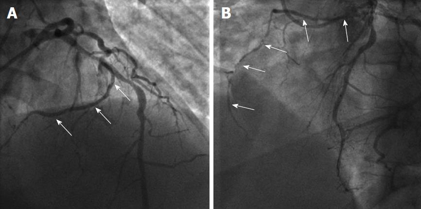
A, B: Angiographic images showing the origin (arrows in A, anteroposterior cranial view) and course (arrows in B, left anterior oblique view) of the right coronary artery from the mid segment of left anterior descending artery. From the authors’ personal dataset and from Ref. [76] (modified with permission).
Figure 12 Single coronary artery from the left sinus.
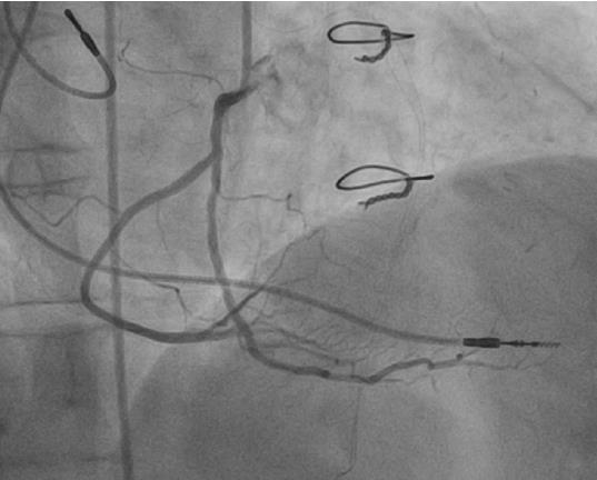
Angiographic images showing the origin and course (arrows) of the aberrant right coronary artery from the distal segment of left circumflex artery (left anterior oblique view). From the authors’ personal dataset.
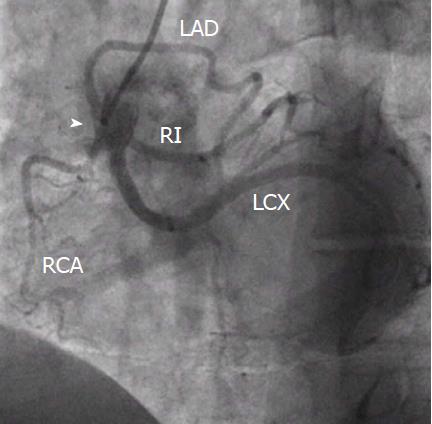
Figure 13 Single coronary artery originating from the right sinus.
Common trunk from the right sinus (arrowhead) providing right coronary artery, left anterior descending artery, left circunflex artery and ramus intermedius. From the authors’ personal dataset. RCA: Right coronary artery; LAD: Left anterior descending; LCX: Left circunflex artery; RI: Ramus intermedius.
Figure 14 Split right coronary artery.
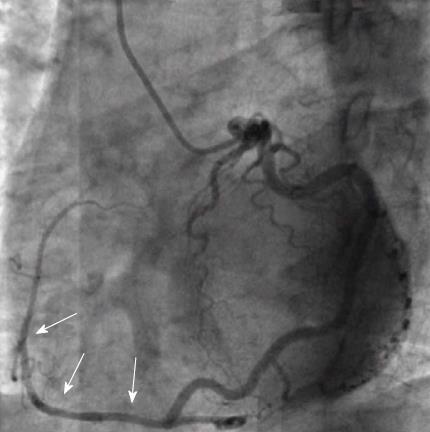
Right coronary artery is divided early into two branches, both of which supply posterior descending arteries. From the authors’ personal dataset.
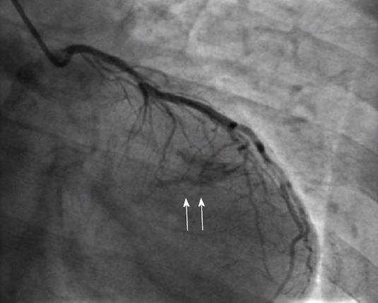
Figure 15 Coronary fistula.
Opacification of the left ventricle during injection of the left coronary artery (arrows) indicate the presence of a fistula. From the authors’ personal dataset.
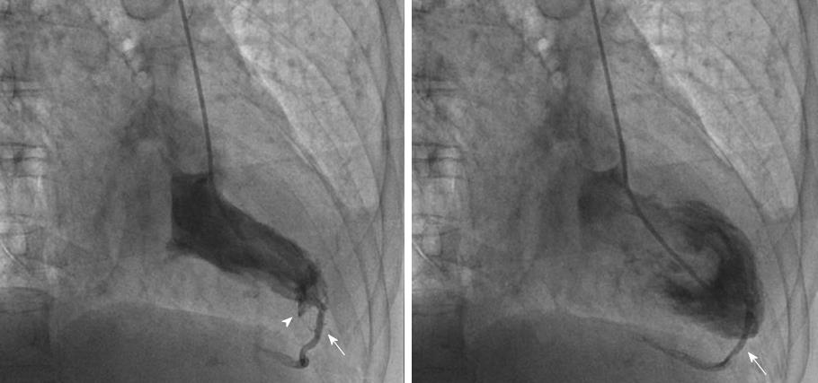
Figure 16 Inadvertent cardiac phlebography through the Thebesian network.
A, B: Right anterior oblique views in systole (A) and diastole (B) during left ventriculography show a minor subendocardial staining (arrowhead) and opacification of a posterior interventricular vein (arrows), due to opening of a 'functional' fistula between the left ventricle and the cardiac vein through the Thebesian network. From the authors’ personal dataset and from Ref. [72] (with permission).
- Citation: Kastellanos S, Aznaouridis K, Vlachopoulos C, Tsiamis E, Oikonomou E, Tousoulis D. Overview of coronary artery variants, aberrations and anomalies. World J Cardiol 2018; 10(10): 127-140
- URL: https://www.wjgnet.com/1949-8462/full/v10/i10/127.htm
- DOI: https://dx.doi.org/10.4330/wjc.v10.i10.127


