Published online May 26, 2014. doi: 10.4331/wjbc.v5.i2.240
Revised: March 13, 2014
Accepted: April 11, 2014
Published online: May 26, 2014
Processing time: 194 Days and 15.3 Hours
AIM: Functional characterization of ATP1A2 mutations that are related to familial or sporadic hemiplegic migraine (FHM2, SHM).
METHODS: cRNA of human Na+/K+-ATPase α2- and β1-subunits were injected in Xenopus laevis oocytes. FHM2 or SHM mutations of residues located in putative α/β interaction sites or in the α2-subunit’s C-terminal region were investigated. Mutants were analyzed by the two-electrode voltage-clamp (TEVC) technique on Xenopus oocytes. Stationary K+-induced Na+/K+ pump currents were measured, and the voltage dependence of apparent K+ affinity was investigated. Transient currents were recorded as ouabain-sensitive currents in Na+ buffers to analyze kinetics and voltage-dependent pre-steady state charge translocations. The expression of constructs was verified by preparation of plasma membrane and total membrane fractions of cRNA-injected oocytes.
RESULTS: Compared to the wild-type enzyme, the mutants G900R and E902K showed no significant differences in the voltage dependence of K+-induced currents, and analysis of the transient currents indicated that the extracellular Na+ affinity was not affected. Mutant G855R showed no pump activity detectable by TEVC. Also for L994del and Y1009X, pump currents could not be recorded. Analysis of the plasma and total membrane fractions showed that the expressed proteins were not or only minimally targeted to the plasma membrane. Whereas the mutation K1003E had no impact on K+ interaction, D999H affected the voltage dependence of K+-induced currents. Furthermore, kinetics of the transient currents was altered compared to the wild-type enzyme, and the apparent affinity for extracellular Na+ was reduced.
CONCLUSION: The investigated FHM2/SHM mutations influence protein function differently depending on the structural impact of the mutated residue.
Core tip: Mutations of the human ATP1A2 gene, which encodes the Na+/K+-ATPase α2-subunit, are associated with familial hemiplegic migraine (FHM2) that is inherited in an autosomal dominant fashion. We studied seven ATP1A2 mutations related to FHM2 or sporadic hemiplegic migraine by electrophysiological and biochemical methods to characterize functional impairments. The mutations G855R, G900R, E902K, L994del, D999H, K1003E and Y1009X were selected according to their structural importance: in putative interaction sites between α- and β-subunit and in the α-subunit’s C-terminal region. Some of these mutations showed a severe loss of function, and we discuss the functional and physiological consequences in order to better understand the molecular basis for neurological impairments.
- Citation: Spiller S, Friedrich T. Functional analysis of human Na+/K+-ATPase familial or sporadic hemiplegic migraine mutations expressed in Xenopus oocytes. World J Biol Chem 2014; 5(2): 240-253
- URL: https://www.wjgnet.com/1949-8454/full/v5/i2/240.htm
- DOI: https://dx.doi.org/10.4331/wjbc.v5.i2.240
Migraine is a common neurological disease, and the different forms are defined by the International Headache Society criteria[1]. Familial hemiplegic migraine (FHM) and sporadic hemiplegic migraine (SHM) are rare autosomal-dominant subforms of migraine with aura. These syndromes are associated with some degree of motor weakness (hemiparesis) and other neurological symptoms during the aura phase. FHM is inherited in an autosomal dominant fashion and genetically heterogeneous. There are a number of mutations related to FHM in three different genes: the CACNA1A gene (FHM1) coding for the neuronal Cav2.1 calcium channel[2,3], the ATP1A2 gene (FHM2) encoding the α2-subunit of the Na+/K+-ATPase[4], and the SCN1A gene (FHM3) encoding the neuronal Nav1.1 sodium channel[5]. The clinical symptoms of SHM are identical to those of FHM but without affected family members.
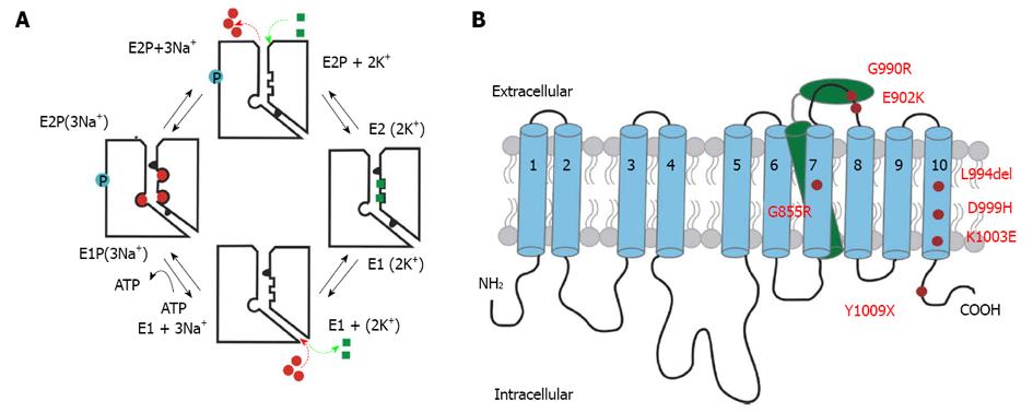
The Na+/K+-ATPase is a transmembrane protein which transports two K+ ions in and three Na+ ions out of the cell upon hydrolysis of ATP (Figure 1A). This electrogenic P-type ATPase assumes two principal conformational changes during its reaction cycle. Upon binding of three intracellular Na+ ions in the ATP-bound E1 conformation, the phosphorylated intermediate with occluded Na+, E1P(3Na+), is formed, followed by a change to the phosphorylated E2P(3Na+) conformation, from which Na+ ions are released to the extracellular medium. Because of the increased affinity for K+ in this configuration, two K+ ions bind subsequently, which triggers the dephosphorylation, and binding of intracellular ATP accelerates the conformational change from E2 to E1. At last, the K+ ions dissociate to the cytoplasm. The sequential translocation of Na+ and K+ ions requires strict cation specificity of the phosphorylation and dephosphorylation reactions. According to the 3Na+/2K+ stoichiometry of transport, electrogenic turnover activity of the Na+/K+-ATPase corresponds to outward movement of one positive charge per reaction cycle, and the major electrogenic event has been shown to take place during extracellular release or reverse binding of Na+[6-8]. This has been suggested to arise from passage of Na+ ions through a narrow, high-field access channel or ‘ion well’[9,10].
The Na+/K+-ATPase consists of at least two mandatory subunits (Figure 1B). The large catalytic α-subunit is composed of ten transmembrane domains (M1-M10), which are linked by five extracellular and four intracellular loops. The smaller regulatory β-subunit is a single-span transmembrane protein (βM) with an ectodomain exhibiting several glycosylation sites. Several isoforms of both subunits are expressed in human cells in a tissue-specific manner. In human brain, the α2-subunit is mainly expressed in glial cells (astrocytes), and loss-of-function of the Na+/K+-ATPase can result in neuronal hyperexcitability, which is commonly explained as follows. The Na+/K+-ATPase maintains the gradients for K+ and Na+ ions, which are essential for the accurate function of secondary active transporters or ion channels, whose activities depend on these gradients. On one hand, changes of the Na+ gradient influence, first, the activity of the Na+/Ca2+ exchanger (NCX) which is crucial for, e.g., Ca2+ signaling. Second, the ability of the glial Na+/glutamate symporter to remove the neurotransmitter glutamate from the synaptic cleft is affected. On the other hand, an altered K+ gradient impairs the repolarizing activity of neuronal K+ channels, which is critical for setting the threshold of action potential generation. Hyperkalemia is known to trigger the phenomenon of cortical spreading depression (CSD), the putatively causal mechanism of the aura phase during a migraine attack[11].
Up to now, far more than 50 mutations of the ATP1A2 gene, which are associated with SHM or FHM2, have been described in literature[12,13]. Yet, most of these mutations have not been studied by electrophysiological techniques, which is a prerequisite for a better understanding of the functional consequences on enzyme activity.
In continuation of previous works[14,15], we studied seven FHM2 or SHM mutations, which are located in regions that are putatively critical for transport properties of the human Na+/K+-ATPase α2-subunit, (Figure 1B), with the two-electrode voltage-clamp technique (TEVC) and biochemical methods to analyze protein expression. Since mutations in the α2-subunit’s C-terminal region were shown to have complex effects on enzyme activity, cation affinities and voltage dependence[16-19], we analyzed four mutations in the transmembrane segment αM10 and in the C-terminus (L994del, K1003E[13], D999H[20] and Y1009X[21]) to further understand structure-function relationships in the C-terminal region. Furthermore, interactions between the α- and β-subunit are not satisfactorily clarified so far. Especially, the highly conserved SYGQ motif in the αM7/M8-loop is believed to interact with the β-ectodomain[22,23]. The FHM2 mutations G900R[24] and E902K[25] are located within this motif and were functionally analyzed in this work. In addition, Gly852 (αM7) has previously been shown to interact with two tyrosines of the βM[26]. In this work, we show that the FHM2 mutation G855R[27] which is located near this interaction site, has severe consequences on the mutant protein’s plasma membrane expression.
As described before[14,19], human Na+/K+-ATPase α2- and β1-subunit cDNAs were subcloned into a modified pCDNA3.1 vector. To distinguish the activity of the heterologously expressed constructs from the endogenous Xenopus Na+/K+-ATPase, the mutations Q116R and N127D were introduced in the human α2-subunit to reduce the ouabain sensitivity (IC50 in a mmol/L range)[28]. This construct is herein referred to as “RD-WT”. Mutants were designed by introducing mutations into the RD-WT α2-construct by site-directed mutagenesis (Quikchange® kit, Stratagene). All PCR-derived fragments were verified by sequencing (Eurofins MWG Operon, Ebersberg, Germany).
cRNA synthesis was carried out with the T7 mMessage mMachine kit (Ambion, Austin, TX). 25 ng of α2- and 2.5 ng of β1-subunit cRNAs were coinjected into oocytes of Xenopus laevis. After three days incubation in ORI buffer (contents in mmol/L: 110 NaCl, 5 KCl, 1 MgCl2, 2 CaCl2, 5 HEPES, pH 7.4, and 50 mg/L gentamycin) at 18 °C, oocytes were subjected to a Na+ loading procedure preceding experiments to elevate [Na+]in. For this purpose, oocytes were incubated for 45 min in Na+ loading solution (contents in mmol/L: 110 NaCl, 2.5 sodium citrate, 5 MOPS, 5 TRIS, pH 7.4) and stored subsequently in Na+ buffer (in mmol/L: 100 NaCl, 1 CaCl2, 5 BaCl2, 2 MgCl2 and 2.5 MOPS, 2.5 TRIS, pH 7.4) for at least 30 min.
Currents were recorded at room temperature (21 °C-23 °C) using a Turbotec 10CX amplifier (NPI instruments, Tamm, Germany) and pClamp 10 software (Axon Inst., Union City, CA). Solutions used for measurements were: Na+ buffer (in mmol/L: 100 NaCl, 1 CaCl2, 5 BaCl2, 2 MgCl2, 2.5 MOPS, 2.5 TRIS, 0.01 ouabain, pH 7.4), and K+ buffers with distinct K+ concentrations, which were prepared by adding appropriate amounts of KCl to Na+ buffer.
K+-induced currents were determined as the difference of currents measured in a distinct K+ buffer and currents measured in Na+ buffer. Oocytes were subjected to the following voltage pulse protocol: from -30 mV holding potential, cells were clamped to potentials between +60 mV and -140 mV (in -20 mV decrements) for 200 ms, followed by a pulse back to -30 mV. All currents within one experiment were normalized to the pump current amplitude at 10 mmol/L K+ and 0 mV. To determine the apparent affinity for extracellular K+, voltage-dependent K0.5(K+ex) values were determined using fits of a Hill equation I = Imax/1+[K0.5/(K+)]nH to the normalized K+-induced currents at a given membrane potential (K0.5 is the concentration at half-maximal current, and nH is the Hill coefficient). nH values from the fits were between 1 and 1.5.
To obtain kinetic information about extracellular Na+ binding/release and the voltage-dependent distribution of pump molecules between E1P and E2P states, pre-steady state currents under Na+/Na+ exchange conditions were recorded. These ouabain-sensitive transient currents were calculated as the difference between currents measured in Na+ buffer with 10 μmol/L ouabain (blocking only the endogenous Na+ pump) and in the presence of 10 mmol/L ouabain (to inhibit the RD-mutated enzyme). Data were fitted by using a monoexponential function, excluding the first 3-5 ms to eliminate capacitive artifacts, yielding time constants τ and amplitudes A. The translocated charge Q was determined from the product A×τ. The resulting Q(V) curves were approximated by a Boltzmann function: Q(V) = Qmin + {(Qmax-Qmin)/1 + exp[zq× F(V-V0.5)/RT]} where Qmax and Qmin are the saturation values of Q(V), V0.5 is the half-maximal voltage at which equal distribution of E1P and E2P states is achieved, zq the fractional charge, F the Faraday constant, R the molar gas constant, T the temperature, and V the membrane potential. After fitting, the translocated charge values were normalized to saturating values (Qmax - Qmin) after subtracting Qmin.
To assess impairments in plasma membrane targeting or expression of mutant proteins that showed no pump current activity in TEVC experiments, plasma membrane (PM) and total membrane (TM) fractions were isolated from oocytes injected with cRNA of the constructs as described before[14,29]. All obtained samples were dissolved in SDS-PAGE sample buffer, and the amount of protein corresponding to the equivalent of two oocytes was separated by 10%SDS-PAGE and blotted on nitrocellulose membranes. Since oocytes are homogenous in size, the procedure of loading the equivalent of a certain number of cells provides an internal loading standard, as shown previously[15]. The α2-subunits of Na+/K+-ATPase were detected with the specific polyclonal antibody AB9094 (Chemicon, Temecula, CA). Afterwards, blots were incubated with a HRP-conjugated secondary antibody (Dako, Glostrup, Denmark). Proteins were visualized by an enhanced chemiluminescence reaction (Roche, Mannheim, Germany).
Structural inspections of the Na+/K+-ATPase (PDB structure entry 3B8E) were carried out with Swiss PDB viewer 3.7. Figures were prepared with PyMOL 1.0r1 (http://www.pymol.org). Data analysis and figure presentation were carried out with Origin 7.0 (OriginLab Corp., Northampton, MA).
Statistical analyses were carried out based on the Student’s t-test for independent samples. The significance level P < 0.05 is indicated in the figures by an “a” above the data points reaching this significance level.
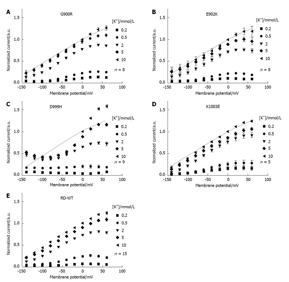
From the investigated ATP1A2 mutants, only G900R, E902K, D999H and K1003E showed K+-induced currents with amplitudes that were sufficiently large for electrophysiological analysis (> 10 nA, Figure 2), whereas no measurable pump activity could be detected for the mutants G855R, L994del and Y1009X. For G900R, E902K and K1003E, the bell-shaped I(V) curves at different [K+]ex did not differ significantly from those of the RD-WT enzyme. This voltage dependence of currents is due to the extracellular competition between K+ and Na+ ions for the two “shared” cation binding sites. With proceeding hyperpolarization of the membrane, reverse binding of extracellular Na+ is stimulated and K+ pump activity inhibited[30,31].
For D999H, in contrast, the voltage dependence of K+-induced currents apparently deviated from RD-WT behavior (Figure 2C). In general, at negative potentials, the current amplitudes of the mutant were small compared to RD-WT amplitudes (data not shown), but at +60 mV, they were in the same range as RD-WT amplitudes (100-200 nA). We suppose that the activity of the D999H construct was similar to the RD-WT enzyme at positive potentials. In contrast to the RD-WT enzyme, the I(V) curves of D999H at high K+ concentrations (2, 5, 10 mmol/L) were nearly constant at potentials between -100 to -40 mV and even increased at hyperpolarization below -100 mV, indicating that the inhibition of K+ pump activity by reverse binding of extracellular Na+ is not as efficient as in the RD-WT enzyme. At potentials more positive than -20 mV, the K+-induced currents started to rise steeply, which shows that positive membrane potentials had a stronger effect on enzyme activity of the D999H mutant compared to the RD-WT enzyme in this voltage range.
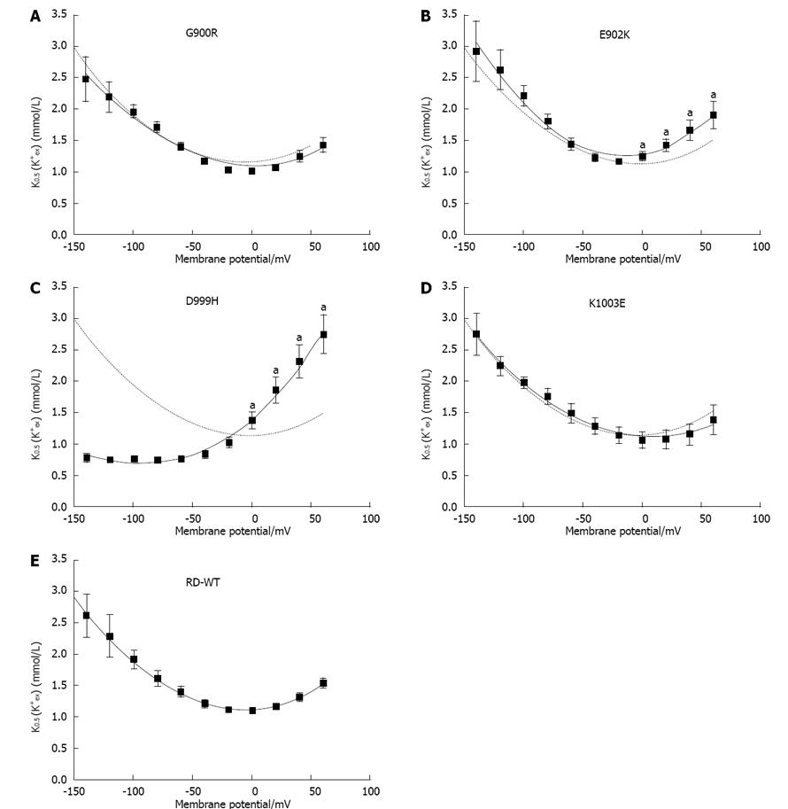
As for the apparent K+ affinity in Na+ containing buffers, K0.5(K+) values were determined from K+-induced currents at different [K+]ex and plotted as a function of the membrane potential (Figure 3). For G900R, E902K, K1003E and RD-WT, the voltage dependence of K0.5(K+) values can be approximated by a parabolic function. The minimal K0.5(K+) values were similar, with values between 1.09-1.25 mmol/L (Table 1). For the RD-WT enzyme, the apparent K+ affinity decreases at negative potentials because the reverse binding of extracellular Na+ is stimulated. In contrast, the K0.5(K+) values determined for mutant D999H did not increase at hyperpolarization, but were nearly voltage-independent between -140 mV and -40 mV (Figure 3C). The minimal K0.5(K+) value was 0.67 mmol/L and shifted to negative potentials. Apparently for D999H, extracellular Na+ does not compete as efficiently with K+ as for the RD-WT enzyme, which indicates a reduced affinity of the mutant for extracellular Na+ (or destabilization of the Na+-bound E2 state). To further investigate this question, the electrogenic Na+/Na+ exchange mode was examined.
| Minimal K0.5 (K+)/mmol/L | Membrane potential at minimum/mV | V0.5/m | zq | |
| RD-WT | 1.12 ± 0.01 | -6.2 ± 1.5 | 0.9 ± 1.3 | 0.77 ± 0.02 |
| G900R | 1.09 ± 0.04 | 0.2 ± 4.7 | 0.3 ± 3.1 | 0.76 ± 0.02 |
| E902K | 1.25 ± 0.03 | -15.2 ± 2.3 | -2.1 ± 2.1 | 0.81 ± 0.02 |
| D999H | 0.67 ± 0.08 | -97.6 ± 4.4 | -67 ± 14 | 0.33 ± 0.11 |
| K1003E | 1.10 ± 0.03 | 6.6 ± 3.3 | -11.3 ± 4.3 | 0.75 ± 0.06 |
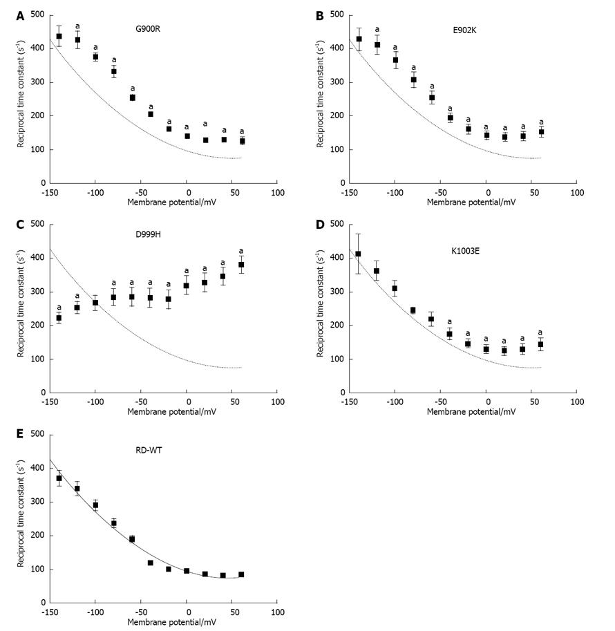
To investigate changes in apparent Na+ex affinity, we measured transient currents under Na+/Na+ exchange conditions (ouabain-sensitive currents, 0 mmol/L K+). Representative transient currents of the RD-WT enzyme are shown as inset in Figure 4E, and the reciprocal time constants of the charge translocation are shown in Figure 5. Basically, the voltage dependence of the reciprocal time constants determined for mutants G900R, E902K and K1003E conformed to that of the RD-WT protein. However, kinetics of charge translocation was slightly faster for these mutants compared to the RD-WT enzyme. Especially for G900R and E902K, the rise of the reciprocal time constants (τ-1) at hyperpolarizing potentials was enhanced.
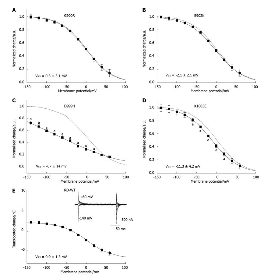
The voltage dependence of charge translocation is shown in Figure 4 and provides information about the distribution of pump molecules between E1P and E2P states[32]. For the mutants G900R and E902K, the Q(V) curves are similar to that of the RD-WT protein, and the V0.5 values in particular did not differ (Table 1). The V0.5 value of mutant K1003E was shifted by -5 to -15 mV. This hints at a slightly reduced apparent Na+ex affinity of this mutant[10,33], which, however, does not seem to impair function in terms of the voltage dependence and the amplitudes of K+-induced currents (Figure 2D).
The D999H mutation had more severe consequences on Na+/Na+ exchange. In general, the transient current signals were fast and small compared to the RD-WT enzyme (data not shown). In addition, the Q(V) curve of translocated charge was linearly dependent on membrane potential, and saturating values were not clearly detectable within the investigated voltage range (Figure 4C). Hence, the approximation with a Boltzmann function and determination of V0.5 proved to be difficult. For fitting, the zq value (Table 1) was reduced until the fitted function superposed the Q values. For this reason, the determined zq can only be regarded as an upper limit, and with a value of 0.33, zq was very small compared to the RD-WT enzyme (0.77). Since V0.5 also directly depends on the quality of the fit, it is likely that the shift of V0.5 by about -70 mV is only a rough estimate for the lower limit of the actual shift. Nonetheless, this strong negative shift shows that D999H has a considerably reduced affinity for extracellular Na+ since very strong hyperpolarization is required to force Na+ ions into the binding sites and to enable the subsequent conformational change to E1P[10,33]. This is in good agreement with the simultaneously reduced K0.5(K+) values at negative potentials. Furthermore, kinetics of the D999H transient currents was less voltage-dependent than for the RD-WT protein (Figure 5C). τ-1 values varied between 200 and 300 s-1 at potentials below 0 mV and increased up to 400 s-1 at depolarization. These results show that the apparent affinities for Na+ and K+ (or stabilization of the cation-occluded state) as well as charge translocation and kinetics of the Na+/Na+ exchange reaction were significantly affected by this mutation.
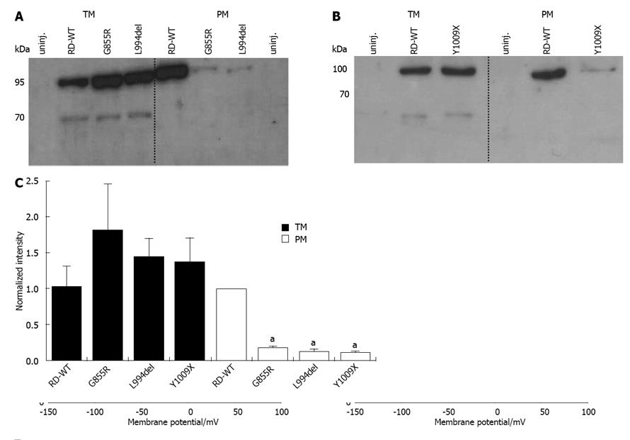
Since the constructs G855R, L994del and Y1009X did not yield measurable Na+/K+ pump currents in TEVC experiments, it was necessary to examine whether or not these proteins were expressed in oocytes and properly targeted to the plasma membrane. For this purpose, plasma membrane (PM) and total intracellular membrane (TM) fractions were prepared using oocytes that had been injected with cRNA of these constructs. Representative Western blots with TM and PM fractions of G855R, L994del (Figure 6C) and Y1009X (Figure 6B) are shown in Figure 6. Densitometric analysis of four Western blots prepared from independent cell batches indicated a disturbed expression pattern of these mutants (Figure 6C). By trend, larger amounts of mutant proteins could be detected in the TM fraction than for the RD-WT protein, which in turn was highly concentrated in the PM fraction. However, analysis of the PM fractions showed that the mutants were not or only minimally expressed in the plasma membrane. The band intensities of PM fractions were only 10%-20% of RD-WT values. Thus, G855R, L994del and Y1009X accumulate in cytoplasmic membranes, and targeting to the plasma membrane was disturbed by these mutations.
α/β-interactions
Several studies have shown that the C-terminal ectodomain of the β-subunit is important for modulation of cation transport by the Na+/K+-ATPase[34-36]. A motif of eight amino acids (Asp897-Tyr905, amino acid sequence DSYGQEWTY) in the αM7/M8-loop seems to be of special importance. Interactions of the β-subunit with this sequence element that encompasses a highly conserved SYGQ motif were identified as crucial for correct folding of newly synthesized α-subunits in the endoplasmic reticulum, and furthermore, it is suspected that an hypothetical sequence motif for proteolytic degradation is masked by these interactions[22,37,38]. Four FHM2/SHM-associated mutations have been identified in the extracellular αM7/M8-loop so far: W887R, G900R, E902K and R908Q[4,24,25,39]. W887R and R908Q, which are not directly located in the SYGQ motif, have already been analyzed[26,40].
The W887R construct was found to be correctly targeted to the plasma membrane of Xenopus oocytes[40], but this mutation caused a complete loss-of-function and a strongly reduced ouabain affinity. Koenderink et al[29] argued that Trp887 might rather have an influence on Arg880, which is critical for ouabain sensitivity, than on targeting-relevant interactions between α- and β-subunits. However, the loss of catalytic function might be due to disturbed α/β-interactions during ion transport. The R908Q mutation, which is very close to the SYGQ motif, indeed affected targeting, since plasma membrane expression in Xenopus oocytes was reduced compared to the RD-WT protein, which easily explains the diminished pump currents[26]. The highly conserved residues Gly900 and Glu902 are located directly in the SYGQ motif and are presumably important for interactions with the β-ectodomain. It was expected that the mutations G900R, which substitutes the small unpolar glycine with a large positively charged arginine, and E902K, where the negatively charged glutamic acid is replaced by a positively charged lysine, would have a strong effect on function. However, both constructs showed no differences compared to the RD-WT enzyme, neither regarding pump activity (Figure 2A, B) nor the apparent affinities for extracellular K+ (K0.5(K+) values in Figure 3A, B) and for extracellular Na+ (Q(V) curves and V0.5 values in Figure 4A, B). Presumably, either these amino acids are not directly interacting with the β-subunit, or the positively charged side chains of arginine and lysine do not interfere with α/β-interactions, at least under the conditions of our study.
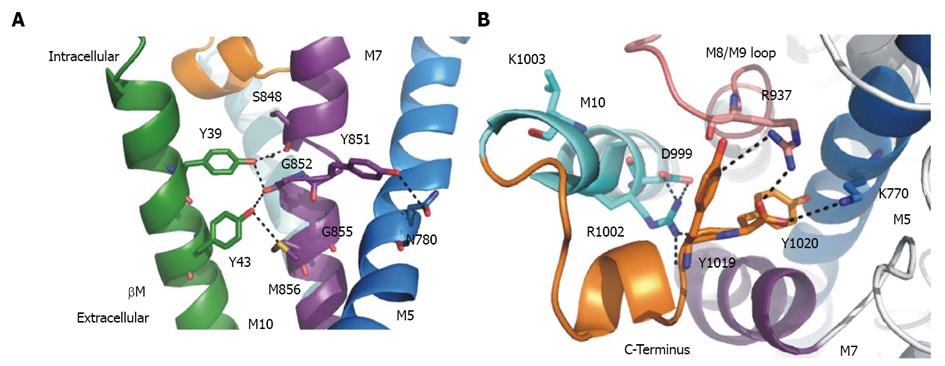
According to the crystal structure of the Na+/K+-ATPase[16,23], Tyr39 and Tyr43 of βM can directly interact with residues at positions 848-856 in αM7 (Figure 7A). Especially, interactions between Gly852 (M7) and both aforementioned tyrosines of the β-subunit seem to stabilize the E2 conformation, and, as confirmed by mutagenesis studies[26,41], not only are hydrogen bonds involved, but also the aromatic ring system of the tyrosines. The β-subunit stabilizes the orientation of αM7 and, consequently, also the position of αM5 because Tyr851 (αM7) can interact with Asn780 in αM5. These interactions are relevant for conformational stabilization during K+ transport[26].
Gly855 is separated by three positions from Gly852, but due to the α-helical structure, it is oriented towards αM5 rather than to βM (Figure 7A). Two mutations at this position have been identified in patients with hemiplegic migraine forms: G855R (FHM2)[27] and G855V (SHM)[13], with G855R presumably having a stronger effect on Na+/K+-ATPase function. Our study indeed shows that the G855R mutant protein is not correctly targeted to the plasma membrane of Xenopus oocytes (Figure 6A, C) although it could well be detected in the total intracellular membrane fraction. However, disruption of α/β-interactions would cause degradation of the protein already in the ER. It is conceivable that the long side chain of the introduced arginine might disturb the structure in a way that transmembrane domains (especially αM7 and αM5) are not correctly positioned. Here, we cannot clarify if the integration in the plasma membrane of G855R is affected because of deficient α/β-interactions or because of misfolding, but Gly855 seems to be a critical position.
In this context, the effect of Y1009X and L994del, which are not targeted to the plasma membrane either but are present in the TM fraction (Figure 6), might be of interest. As shown in Figure 7A, the flexible C-terminus (orange) of the α-subunit is oriented towards a region between βM and αM7, in interaction distance to Lys770 in αM5 (Figure 7B). It was suggested that Tyr998 in αM10 directly interacts with βM[23]. The Y1009X mutant protein lacks the 11 C-terminal residues, and in L994del, the 25 C-terminal amino acid residues are shifted N-terminally by one position. These modifications in the C-terminus might affect the orientation of αM7 and αM5 and thereby, correct protein folding. To what extent α/β-interactions are influenced cannot be clarified in this study.
A number of functional studies imply that the C-terminus is intimately involved in the stabilization of the third Na+ binding site[16,19,42,43], including analyses of mutations which are suspected to trigger neurological diseases. Elongation of the C-terminus provoked different functional abnormalities. Investigations on a mutation found in a patient with rapid-onset dystonia parkinsonism, where the α3-subunit’s C-terminus is extended by one tyrosine, implied a direct participation of the C-terminus in Na+ binding[43]. Another C-terminal mutation X1021R (mutation of the stop codon resulting in an elongation of the C-terminus by 28 amino acids) was analyzed electrophysiologically in Xenopus oocytes[14]. Interestingly, this mutation affected the apparent Na+ex affinity of the enzyme in a similar way as the D999H mutation. The Q(V) curve of transient currents of X1021R was comparably shallow, as for D999H (Figure 4C), and linear in the tested potential range. The zq value was reduced to 0.3 for both mutations, which implies that Na+ release and rebinding is less voltage-dependent. Furthermore, the reciprocal time constants of transient currents showed inverse voltage dependence compared to the RD-WT enzyme (kinetics accelerated with increasing potentials, Figure 5C). Similar curves were also detected for other C-terminally mutated enzymes like ΔYY or ΔKE(S/T)YY (deletion of the last two or five amino acids, depending on species isoform)[17-19]. The transient currents correlate with the movement of the third Na+ ion through a substantial fraction of the membrane dielectric, which reaches its bindings site through a high-field access channel[10,33]. The τ-1(V) curve measured for D999H (Figure 5C) or ΔYY[19] corresponds to a voltage dependence that is predicted by Vasilyev et al[19,44] for a reaction cycle in which the intra- and extracellular access for Na+ to its binding sites is facilitated. In conclusion, the C-terminus stabilizes the Na+-occluded state. This argumentation is also shared by Vedovato and Gadsby, who argued that the C-terminally deleted mutations increase the free energy for E1P(3Na+)[18]. This destabilization manifests in a faster conformational change or in a faster access/release of intracellular Na+ ions, which means that the function of the E1P(3Na+) state is impaired and correct closure of an intracellular occlusion gate for Na+ ions is not assured.
Not only are the two terminal tyrosines involved in this stabilization, but also the residues Arg937 (αM8/M9-loop), Asp999 (M10) and Arg1002 (M10) are part of a network of interactions with these tyrosines (Figure 7B). The FHM2/SHM mutations R937P, R1002Q[42] and D999H, as well as the ΔYY or ΔKE(S/T)YY sequence variants have similar effects on transient currents (kinetics and Q(V) distribution). The functional studies all show that the C-terminus not only regulates the apparent Na+ex affinity in the E2P conformation, but also the Na+in affinity in the E1 conformation[17,19,43]. Based on molecular dynamics simulations of the wild-type enzyme and C-terminally mutated α2-subunits, it was proposed that the amino acids Arg937, Asp999, Arg1002 and Tyr1019/1020 form an intracellular ion pathway with Asp930 at its end, which controls the access to the third Na+ binding site depending on the protonation state of Asp930[42]. Our study confirms that Asp999 is at least indirectly involved in the stabilization of Na+ binding because its substitution by a histidine affected electrogenicity and kinetics of Na+ charge translocation in a similar fashion. In contrast, the overall electrophysiological data of K1003E did not show severe functional abnormalities, and with regard to the crystal structure of the Na+/K+-ATPase, we conclude that Lys1003 (αM10) is not directly involved in the C-terminal network (Figure 7B).
Dysfunction of the Na+/K+-ATPase affects excitatory processes in the CNS, especially in patients suffering from hemiplegic migraine. How do the mutations studied in this work affect the physiological processes in neuronal signaling cascades, since the α2-isoform in human brain is mainly expressed in astrocytes and not in neurons? The CSD phenomenon is discussed as pathophysiological mechanism of the migraine aura. It is promoted by hyperexcitability caused by insufficient removal of K+ and neurotransmitters such as glutamate from the synaptic cleft, which is the primary function of astrocytes. The glial Na+/K+-ATPase is directly involved in K+ transport, and it indirectly influences glutamate and Ca2+ transport by regulating the Na+ gradient, which is the energy source of the glutamate transporter (EAAT) and the Na+/Ca2+-exchanger (NCX).
G900R, E902K and K1003E did not show significant functional abnormalities compared to the RD-WT enzyme, at least under the conditions tested here. It is possible that these mutations impair the enzymatic function in human cells e.g. due to different temperature conditions (37 °C as opposed to oocytes, which need to be kept at room temperature), as shown previously for another FHM2 mutation P979L[15]. Furthermore, the constructs G855R, L994del and Y1009X exhibited strongly reduced expression in the plasma membrane (Figure 6). This hints at an incomplete or improper folding of the protein so that these mutants could not be correctly targeted to the plasma membrane. In patients with such mutations, the pump enzyme is seriously damaged, and cannot contribute to the maintenance of ion gradients or to the removal of K+. As a consequence, hyperexcitability is probable.
Compared to all other mutants in this study, which gave rise to measurable Na+/K+ pump currents, the D999H mutation had the largest impact on pump function. The voltage dependence of Na+/K+ pump activity was shifted to positive potentials compared to the RD-WT enzyme (Figure 2C). We suppose that K+ transport of this construct is only effective at around zero or positive membrane potentials. Since the α2-isoform is dominant in astrocytes with resting potentials at -85 to -90 mV, this mutant exhibits a severe loss-of-function. K+ cannot be removed efficiently from the synaptic cleft at negative potentials, which lowers the excitation threshold and may trigger CSD. Furthermore, regarding the negative shift of the Q(V) curve (Na+/Na+ exchange conditions, Figure 4C) and the low K0.5(K+) values at hyperpolarization (Figure 3C), we conclude that the apparent affinity for extracellular Na+ is reduced in the D999H mutant. As explained above, Asp999 is part of the C-terminal interaction network which plays a role in Na+ binding (especially concerning stabilization of the third Na+ binding site, Figure 7B). Mutations at positions Arg937 and Tyr1019/Tyr1020, which are also part of this network, affected the affinity for both, intra- and extracellular Na+[17,19,43]. The ATP1A2 α2-isoform (expressed in non-excitable cells of the CNS) has a slightly increased Na+in affinity compared to the α3-isoform[45,46], which is expressed in neurons. This is advantageous because enzyme activity in astrocytes presumably depends mainly on the increase of the intracellular Na+ concentration. In other words, [Na+]in is the important factor determining the sensitivity of the Na+/K+-ATPase towards increasing extracellular K+[47]. For instance, the intracellular Na+ concentration increases upon glutamate uptake by EAAT, and this stimulates pump activity and K+ transport. Accordingly, a reduced Na+in affinity would constrain forward pumping with serious consequences for the recovery of the neuronal resting potential.
In effect, K+ and glutamate removal from the synaptic cleft not only depends on Na+/K+-ATPase activity, but other transporting enzymes are also involved. Furthermore, the penetrance of ATP1A2 mutations can be low or heterogenous because of a large diversity of phenotypic expression depending on genetic and environmental conditions[48-50]. In consequence, physiological impacts of α2-mutations vary and provoke clinical symptoms of different severity.
In conclusion, this study shows that the investigated FHM2/SHM mutations influence protein function differently depending on the structural impacts of the mutated residue, and thereby, the spectrum of molecular phenotypes of ATP1A2 mutations is widened. We have identified at least two positions that are critical for correct protein function, with Asp999 being involved in Na+-binding and with Gly855 being essential for plasma membrane targeting. The functional analysis of FHM2/SHM mutations are mandatory to elucidate structure-function relationships of the Na+/K+-ATPase and, furthermore, to identify biochemical linkage between impairments of protein function and neurological diseases. Our results may help to understand molecular mechanisms in order to develop a basic approach for future therapeutic strategies.
The authors thank Dr. Neslihan Tavraz and Dr. Kirstin Hobiger for valuable discussions; and the German Research Foundation (Cluster of Excellence “Unifying Concepts in Catalysis”) for financial support.
The Na+/K+-ATPase is a very important transmembrane protein in the signaling cascade and it has been investigated for over 50 years. There are still open questions concerning details of the reaction mechanism and structure-function relationships. In patients suffering from a genetically inherited subform of migraine with aura (familial hemiplegic migraine), mutations of the ATP1A2 gene, which codes for the α2-subunit of the Na+/K+-ATPase, have been identified.
To clarify structure-function relationships of the Na+/K+-ATPase, different methods have to be applied like molecular dynamics simulations, crystallography, mutagenesis studies together with biochemical assays or electrophysiology. Especially, interactions between the two mandatory enzyme subunits, the role of the α-subunit’s C-terminus and the detailed mechanism of Na+ binding remain unclear. This study analyzed ATP1A2 mutants functionally by electrophysiological and biochemical methods to clarify some of these questions.
More than 50 mutations of the ATP1A2 gene associated with familial hemiplegic migraine have been identified, but many of them have not been functionally analyzed. This study identifies critical structure elements of the Na+/K+-ATPase and discusses their impact on correct protein function. After publication of the first crystal structure, many efforts were made to clarify the role of the α-subunit’s C-terminus and its structural interaction. The authors show in this study that Asp999 is indeed part of the C-terminal network and is critical for Na+ binding. Furthermore, the authors have identified Gly855 to be a very critical position for correct protein function.
This study helps to elucidate structure-function relationships of the Na+/K+-ATPase and its correlation with neurological diseases. It is mandatory to understand the molecular basis of genotype-phenotype relations and to develop therapeutic approaches and future therapeutic strategies.
The Na+/K+-ATPase is an ion pump. This transmembrane protein maintains the electrochemical gradients for sodium and potassium ions, which are necessary for the transmission of stimuli in neurons or muscle cells. The Na+/K+-ATPase can be inhibited by ouabain, a cardiac glycoside which was used for the treatment of heart diseases. Xenopus oocytes are the eggs of the African Clawed Frog. They are used for the expression of proteins like ion channels or ion pumps to study ion transport by electrophysiological methods. The two-electrode voltage-clamp technique is used to measure changes in conductivity and ion currents over the cell membrane. With this method, it is possible to control the membrane potential of the cell and to analyze current-voltage relationships of ion-transporting membrane proteins.
This paper represent a very good piece of scientific information, it provides information on the consequences of mutations in the Na,K-ATPase alpha subunit, measured by voltage clamp.
P- Reviewers: Grover AK, Lesage F, Mahmmoud YA, Trumper L S- Editor: Song XX L- Editor: A E- Editor: Lu YJ
| 1. | Headache Classification Subcommittee of the International Headache Society. The International Classification of Headache Disorders: 2nd edition. Cephalalgia. 2004;24 Suppl 1:9-160. [RCA] [PubMed] [DOI] [Full Text] [Cited by in Crossref: 798] [Cited by in RCA: 2427] [Article Influence: 115.6] [Reference Citation Analysis (0)] |
| 2. | Ophoff RA, Terwindt GM, Vergouwe MN, van Eijk R, Oefner PJ, Hoffman SM, Lamerdin JE, Mohrenweiser HW, Bulman DE, Ferrari M. Familial hemiplegic migraine and episodic ataxia type-2 are caused by mutations in the Ca2+ channel gene CACNL1A4. Cell. 1996;87:543-552. [RCA] [PubMed] [DOI] [Full Text] [Cited by in Crossref: 1615] [Cited by in RCA: 1511] [Article Influence: 52.1] [Reference Citation Analysis (0)] |
| 3. | Ducros A, Denier C, Joutel A, Cecillon M, Lescoat C, Vahedi K, Darcel F, Vicaut E, Bousser MG, Tournier-Lasserve E. The clinical spectrum of familial hemiplegic migraine associated with mutations in a neuronal calcium channel. N Engl J Med. 2001;345:17-24. [RCA] [PubMed] [DOI] [Full Text] [Cited by in Crossref: 400] [Cited by in RCA: 364] [Article Influence: 15.2] [Reference Citation Analysis (0)] |
| 4. | De Fusco M, Marconi R, Silvestri L, Atorino L, Rampoldi L, Morgante L, Ballabio A, Aridon P, Casari G. Haploinsufficiency of ATP1A2 encoding the Na+/K+ pump alpha2 subunit associated with familial hemiplegic migraine type 2. Nat Genet. 2003;33:192-196. [RCA] [PubMed] [DOI] [Full Text] [Cited by in Crossref: 703] [Cited by in RCA: 648] [Article Influence: 29.5] [Reference Citation Analysis (0)] |
| 5. | Dichgans M, Freilinger T, Eckstein G, Babini E, Lorenz-Depiereux B, Biskup S, Ferrari MD, Herzog J, van den Maagdenberg AM, Pusch M. Mutation in the neuronal voltage-gated sodium channel SCN1A in familial hemiplegic migraine. Lancet. 2005;366:371-377. [RCA] [PubMed] [DOI] [Full Text] [Cited by in Crossref: 614] [Cited by in RCA: 566] [Article Influence: 28.3] [Reference Citation Analysis (0)] |
| 6. | Fendler K, Grell E, Haubs M, Bamberg E. Pump currents generated by the purified Na+K+-ATPase from kidney on black lipid membranes. EMBO J. 1985;4:3079-3085. [PubMed] |
| 7. | Nakao M, Gadsby DC. Voltage dependence of Na translocation by the Na/K pump. Nature. 1986;323:628-630. [RCA] [PubMed] [DOI] [Full Text] [Cited by in Crossref: 178] [Cited by in RCA: 184] [Article Influence: 4.7] [Reference Citation Analysis (0)] |
| 8. | Wuddel I, Apell HJ. Electrogenicity of the sodium transport pathway in the Na,K-ATPase probed by charge-pulse experiments. Biophys J. 1995;69:909-921. [RCA] [PubMed] [DOI] [Full Text] [Cited by in Crossref: 86] [Cited by in RCA: 81] [Article Influence: 2.7] [Reference Citation Analysis (0)] |
| 9. | Sagar A, Rakowski RF. Access channel model for the voltage dependence of the forward-running Na+/K+ pump. J Gen Physiol. 1994;103:869-893. [RCA] [PubMed] [DOI] [Full Text] [Full Text (PDF)] [Cited by in Crossref: 68] [Cited by in RCA: 79] [Article Influence: 2.5] [Reference Citation Analysis (0)] |
| 10. | Holmgren M, Wagg J, Bezanilla F, Rakowski RF, De Weer P, Gadsby DC. Three distinct and sequential steps in the release of sodium ions by the Na+/K+-ATPase. Nature. 2000;403:898-901. [RCA] [PubMed] [DOI] [Full Text] [Cited by in Crossref: 132] [Cited by in RCA: 122] [Article Influence: 4.9] [Reference Citation Analysis (0)] |
| 11. | Eikermann-Haerter K, Negro A, Ayata C. Spreading depression and the clinical correlates of migraine. Rev Neurosci. 2013;24:353-363. [RCA] [PubMed] [DOI] [Full Text] [Cited by in Crossref: 27] [Cited by in RCA: 33] [Article Influence: 2.8] [Reference Citation Analysis (0)] |
| 12. | Morth JP, Poulsen H, Toustrup-Jensen MS, Schack VR, Egebjerg J, Andersen JP, Vilsen B, Nissen P. The structure of the Na+,K+-ATPase and mapping of isoform differences and disease-related mutations. Philos Trans R Soc Lond B Biol Sci. 2009;364:217-227. [RCA] [PubMed] [DOI] [Full Text] [Cited by in Crossref: 67] [Cited by in RCA: 67] [Article Influence: 4.2] [Reference Citation Analysis (0)] |
| 13. | Riant F, Ducros A, Ploton C, Barbance C, Depienne C, Tournier-Lasserve E. De novo mutations in ATP1A2 and CACNA1A are frequent in early-onset sporadic hemiplegic migraine. Neurology. 2010;75:967-972. [RCA] [PubMed] [DOI] [Full Text] [Cited by in Crossref: 95] [Cited by in RCA: 92] [Article Influence: 6.1] [Reference Citation Analysis (0)] |
| 14. | Tavraz NN, Friedrich T, Dürr KL, Koenderink JB, Bamberg E, Freilinger T, Dichgans M. Diverse functional consequences of mutations in the Na+/K+-ATPase alpha2-subunit causing familial hemiplegic migraine type 2. J Biol Chem. 2008;283:31097-31106. [RCA] [PubMed] [DOI] [Full Text] [Cited by in Crossref: 66] [Cited by in RCA: 71] [Article Influence: 4.2] [Reference Citation Analysis (0)] |
| 15. | Tavraz NN, Dürr KL, Koenderink JB, Freilinger T, Bamberg E, Dichgans M, Friedrich T. Impaired plasma membrane targeting or protein stability by certain ATP1A2 mutations identified in sporadic or familial hemiplegic migraine. Channels (Austin). 2009;3:82-87. [RCA] [PubMed] [DOI] [Full Text] [Cited by in Crossref: 26] [Cited by in RCA: 33] [Article Influence: 2.1] [Reference Citation Analysis (0)] |
| 16. | Morth JP, Pedersen BP, Toustrup-Jensen MS, Sørensen TL, Petersen J, Andersen JP, Vilsen B, Nissen P. Crystal structure of the sodium-potassium pump. Nature. 2007;450:1043-1049. [RCA] [PubMed] [DOI] [Full Text] [Cited by in Crossref: 687] [Cited by in RCA: 663] [Article Influence: 39.0] [Reference Citation Analysis (0)] |
| 17. | Yaragatupalli S, Olivera JF, Gatto C, Artigas P. Altered Na+ transport after an intracellular alpha-subunit deletion reveals strict external sequential release of Na+ from the Na/K pump. Proc Natl Acad Sci USA. 2009;106:15507-15512. [RCA] [PubMed] [DOI] [Full Text] [Cited by in Crossref: 38] [Cited by in RCA: 35] [Article Influence: 2.2] [Reference Citation Analysis (0)] |
| 18. | Vedovato N, Gadsby DC. The two C-terminal tyrosines stabilize occluded Na/K pump conformations containing Na or K ions. J Gen Physiol. 2010;136:63-82. [RCA] [PubMed] [DOI] [Full Text] [Full Text (PDF)] [Cited by in Crossref: 27] [Cited by in RCA: 26] [Article Influence: 1.7] [Reference Citation Analysis (0)] |
| 19. | Meier S, Tavraz NN, Dürr KL, Friedrich T. Hyperpolarization-activated inward leakage currents caused by deletion or mutation of carboxy-terminal tyrosines of the Na+/K+-ATPase {alpha} subunit. J Gen Physiol. 2010;135:115-134. [RCA] [PubMed] [DOI] [Full Text] [Full Text (PDF)] [Cited by in Crossref: 39] [Cited by in RCA: 34] [Article Influence: 2.3] [Reference Citation Analysis (0)] |
| 20. | Fernandez DM, Hand CK, Sweeney BJ, Parfrey NA. A novel ATP1A2 gene mutation in an Irish familial hemiplegic migraine kindred. Headache. 2008;48:101-108. [RCA] [PubMed] [DOI] [Full Text] [Cited by in Crossref: 4] [Cited by in RCA: 13] [Article Influence: 0.8] [Reference Citation Analysis (0)] |
| 21. | Gallanti A, Tonelli A, Cardin V, Bussone G, Bresolin N, Bassi MT. A novel de novo nonsense mutation in ATP1A2 associated with sporadic hemiplegic migraine and epileptic seizures. J Neurol Sci. 2008;273:123-126. [RCA] [PubMed] [DOI] [Full Text] [Cited by in Crossref: 26] [Cited by in RCA: 32] [Article Influence: 1.9] [Reference Citation Analysis (0)] |
| 22. | Béguin P, Hasler U, Staub O, Geering K. Endoplasmic reticulum quality control of oligomeric membrane proteins: topogenic determinants involved in the degradation of the unassembled Na,K-ATPase alpha subunit and in its stabilization by beta subunit assembly. Mol Biol Cell. 2000;11:1657-1672. [RCA] [PubMed] [DOI] [Full Text] [Cited by in Crossref: 40] [Cited by in RCA: 45] [Article Influence: 1.8] [Reference Citation Analysis (0)] |
| 23. | Shinoda T, Ogawa H, Cornelius F, Toyoshima C. Crystal structure of the sodium-potassium pump at 2.4 A resolution. Nature. 2009;459:446-450. [RCA] [PubMed] [DOI] [Full Text] [Cited by in Crossref: 498] [Cited by in RCA: 475] [Article Influence: 29.7] [Reference Citation Analysis (0)] |
| 24. | Deprez L, Peeters K, Van Paesschen W, Claeys KG, Claes LR, Suls A, Audenaert D, Van Dyck T, Goossens D, Del-Favero J. Familial occipitotemporal lobe epilepsy and migraine with visual aura: linkage to chromosome 9q. Neurology. 2007;68:1995-2002. [RCA] [PubMed] [DOI] [Full Text] [Cited by in Crossref: 1] [Cited by in RCA: 2] [Article Influence: 0.1] [Reference Citation Analysis (0)] |
| 25. | Jurkat-Rott K, Freilinger T, Dreier JP, Herzog J, Göbel H, Petzold GC, Montagna P, Gasser T, Lehmann-Horn F, Dichgans M. Variability of familial hemiplegic migraine with novel A1A2 Na+/K+-ATPase variants. Neurology. 2004;62:1857-1861. [RCA] [PubMed] [DOI] [Full Text] [Cited by in Crossref: 112] [Cited by in RCA: 117] [Article Influence: 5.6] [Reference Citation Analysis (0)] |
| 26. | Dürr KL, Tavraz NN, Dempski RE, Bamberg E, Friedrich T. Functional significance of E2 state stabilization by specific alpha/beta-subunit interactions of Na,K- and H,K-ATPase. J Biol Chem. 2009;284:3842-3854. [RCA] [PubMed] [DOI] [Full Text] [Cited by in Crossref: 23] [Cited by in RCA: 25] [Article Influence: 1.5] [Reference Citation Analysis (0)] |
| 27. | de Vries B, Stam AH, Kirkpatrick M, Vanmolkot KR, Koenderink JB, van den Heuvel JJ, Stunnenberg B, Goudie D, Shetty J, Jain V. Familial hemiplegic migraine is associated with febrile seizures in an FHM2 family with a novel de novo ATP1A2 mutation. Epilepsia. 2009;50:2503-2504. [RCA] [PubMed] [DOI] [Full Text] [Cited by in Crossref: 18] [Cited by in RCA: 19] [Article Influence: 1.2] [Reference Citation Analysis (0)] |
| 28. | Price EM, Lingrel JB. Structure-function relationships in the Na,K-ATPase alpha subunit: site-directed mutagenesis of glutamine-111 to arginine and asparagine-122 to aspartic acid generates a ouabain-resistant enzyme. Biochemistry. 1988;27:8400-8408. [RCA] [PubMed] [DOI] [Full Text] [Cited by in Crossref: 330] [Cited by in RCA: 329] [Article Influence: 8.9] [Reference Citation Analysis (0)] |
| 29. | Koenderink JB, Geibel S, Grabsch E, De Pont JJ, Bamberg E, Friedrich T. Electrophysiological analysis of the mutated Na,K-ATPase cation binding pocket. J Biol Chem. 2003;278:51213-51222. [RCA] [PubMed] [DOI] [Full Text] [Cited by in Crossref: 50] [Cited by in RCA: 53] [Article Influence: 2.4] [Reference Citation Analysis (0)] |
| 30. | Nakao M, Gadsby DC. [Na] and [K] dependence of the Na/K pump current-voltage relationship in guinea pig ventricular myocytes. J Gen Physiol. 1989;94:539-565. [RCA] [PubMed] [DOI] [Full Text] [Full Text (PDF)] [Cited by in Crossref: 171] [Cited by in RCA: 198] [Article Influence: 5.5] [Reference Citation Analysis (0)] |
| 31. | Rakowski RF, Gadsby DC, De Weer P. Stoichiometry and voltage dependence of the sodium pump in voltage-clamped, internally dialyzed squid giant axon. J Gen Physiol. 1989;93:903-941. [RCA] [PubMed] [DOI] [Full Text] [Full Text (PDF)] [Cited by in Crossref: 118] [Cited by in RCA: 134] [Article Influence: 3.7] [Reference Citation Analysis (0)] |
| 32. | Rakowski RF. Charge movement by the Na/K pump in Xenopus oocytes. J Gen Physiol. 1993;101:117-144. [RCA] [PubMed] [DOI] [Full Text] [Full Text (PDF)] [Cited by in Crossref: 77] [Cited by in RCA: 83] [Article Influence: 2.6] [Reference Citation Analysis (0)] |
| 33. | Holmgren M, Rakowski RF. Charge translocation by the Na+/K+ pump under Na+/Na+ exchange conditions: intracellular Na+ dependence. Biophys J. 2006;90:1607-1616. [RCA] [PubMed] [DOI] [Full Text] [Cited by in Crossref: 30] [Cited by in RCA: 30] [Article Influence: 1.6] [Reference Citation Analysis (0)] |
| 34. | Jaunin P, Jaisser F, Beggah AT, Takeyasu K, Mangeat P, Rossier BC, Horisberger JD, Geering K. Role of the transmembrane and extracytoplasmic domain of beta subunits in subunit assembly, intracellular transport, and functional expression of Na,K-pumps. J Cell Biol. 1993;123:1751-1759. [RCA] [PubMed] [DOI] [Full Text] [Full Text (PDF)] [Cited by in Crossref: 66] [Cited by in RCA: 69] [Article Influence: 2.2] [Reference Citation Analysis (0)] |
| 35. | Hasler U, Wang X, Crambert G, Béguin P, Jaisser F, Horisberger JD, Geering K. Role of beta-subunit domains in the assembly, stable expression, intracellular routing, and functional properties of Na,K-ATPase. J Biol Chem. 1998;273:30826-30835. [RCA] [PubMed] [DOI] [Full Text] [Cited by in Crossref: 116] [Cited by in RCA: 120] [Article Influence: 4.4] [Reference Citation Analysis (0)] |
| 36. | Eakle KA, Kabalin MA, Wang SG, Farley RA. The influence of beta subunit structure on the stability of Na+/K(+)-ATPase complexes and interaction with K+. J Biol Chem. 1994;269:6550-6557. [PubMed] |
| 37. | Colonna TE, Huynh L, Fambrough DM. Subunit interactions in the Na,K-ATPase explored with the yeast two-hybrid system. J Biol Chem. 1997;272:12366-12372. [RCA] [PubMed] [DOI] [Full Text] [Cited by in Crossref: 77] [Cited by in RCA: 78] [Article Influence: 2.8] [Reference Citation Analysis (0)] |
| 38. | Geering K. The functional role of beta subunits in oligomeric P-type ATPases. J Bioenerg Biomembr. 2001;33:425-438. [PubMed] |
| 39. | de Vries B, Freilinger T, Vanmolkot KR, Koenderink JB, Stam AH, Terwindt GM, Babini E, van den Boogerd EH, van den Heuvel JJ, Frants RR. Systematic analysis of three FHM genes in 39 sporadic patients with hemiplegic migraine. Neurology. 2007;69:2170-2176. [RCA] [PubMed] [DOI] [Full Text] [Cited by in Crossref: 96] [Cited by in RCA: 74] [Article Influence: 4.1] [Reference Citation Analysis (0)] |
| 40. | Koenderink JB, Zifarelli G, Qiu LY, Schwarz W, De Pont JJ, Bamberg E, Friedrich T. Na,K-ATPase mutations in familial hemiplegic migraine lead to functional inactivation. Biochim Biophys Acta. 2005;1669:61-68. [RCA] [PubMed] [DOI] [Full Text] [Cited by in Crossref: 43] [Cited by in RCA: 46] [Article Influence: 2.3] [Reference Citation Analysis (0)] |
| 41. | Hasler U, Crambert G, Horisberger JD, Geering K. Structural and functional features of the transmembrane domain of the Na,K-ATPase beta subunit revealed by tryptophan scanning. J Biol Chem. 2001;276:16356-16364. [RCA] [PubMed] [DOI] [Full Text] [Cited by in Crossref: 48] [Cited by in RCA: 51] [Article Influence: 2.1] [Reference Citation Analysis (0)] |
| 42. | Poulsen H, Khandelia H, Morth JP, Bublitz M, Mouritsen OG, Egebjerg J, Nissen P. Neurological disease mutations compromise a C-terminal ion pathway in the Na(+)/K(+)-ATPase. Nature. 2010;467:99-102. [RCA] [PubMed] [DOI] [Full Text] [Cited by in Crossref: 104] [Cited by in RCA: 100] [Article Influence: 6.7] [Reference Citation Analysis (0)] |
| 43. | Blanco-Arias P, Einholm AP, Mamsa H, Concheiro C, Gutiérrez-de-Terán H, Romero J, Toustrup-Jensen MS, Carracedo A, Jen JC, Vilsen B. A C-terminal mutation of ATP1A3 underscores the crucial role of sodium affinity in the pathophysiology of rapid-onset dystonia-parkinsonism. Hum Mol Genet. 2009;18:2370-2377. [RCA] [PubMed] [DOI] [Full Text] [Cited by in Crossref: 50] [Cited by in RCA: 51] [Article Influence: 3.2] [Reference Citation Analysis (0)] |
| 44. | Vasilyev A, Khater K, Rakowski RF. Effect of extracellular pH on presteady-state and steady-state current mediated by the Na+/K+ pump. J Membr Biol. 2004;198:65-76. [RCA] [PubMed] [DOI] [Full Text] [Cited by in Crossref: 17] [Cited by in RCA: 18] [Article Influence: 0.9] [Reference Citation Analysis (0)] |
| 45. | Blanco G, Mercer RW. Isozymes of the Na-K-ATPase: heterogeneity in structure, diversity in function. Am J Physiol. 1998;275:F633-F650. [PubMed] |
| 46. | Crambert G, Hasler U, Beggah AT, Yu C, Modyanov NN, Horisberger JD, Lelièvre L, Geering K. Transport and pharmacological properties of nine different human Na, K-ATPase isozymes. J Biol Chem. 2000;275:1976-1986. [RCA] [PubMed] [DOI] [Full Text] [Cited by in Crossref: 332] [Cited by in RCA: 331] [Article Influence: 13.2] [Reference Citation Analysis (0)] |
| 47. | Rose CR, Ransom BR. Gap junctions equalize intracellular Na+ concentration in astrocytes. Glia. 1997;20:299-307. [RCA] [PubMed] [DOI] [Full Text] [Cited by in RCA: 1] [Reference Citation Analysis (0)] |
| 48. | Ducros A, Joutel A, Vahedi K, Cecillon M, Ferreira A, Bernard E, Verier A, Echenne B, Lopez de Munain A, Bousser MG. Mapping of a second locus for familial hemiplegic migraine to 1q21-q23 and evidence of further heterogeneity. Ann Neurol. 1997;42:885-890. [RCA] [PubMed] [DOI] [Full Text] [Cited by in Crossref: 143] [Cited by in RCA: 117] [Article Influence: 4.2] [Reference Citation Analysis (0)] |
| 49. | Haan J, Terwindt GM, van den Maagdenberg AM, Stam AH, Ferrari MD. A review of the genetic relation between migraine and epilepsy. Cephalalgia. 2008;28:105-113. [RCA] [PubMed] [DOI] [Full Text] [Cited by in RCA: 14] [Reference Citation Analysis (0)] |
| 50. | Shyti R, de Vries B, van den Maagdenberg A. Migraine genes and the relation to gender. Headache. 2011;51:880-890. [RCA] [PubMed] [DOI] [Full Text] [Cited by in Crossref: 29] [Cited by in RCA: 32] [Article Influence: 2.7] [Reference Citation Analysis (0)] |


