Editorial Open Access
Copyright ©2011 Baishideng Publishing Group Co., Limited. All rights reserved.
World J Biol Chem. Sep 26, 2011; 2(9): 193-201
Published online Sep 26, 2011. doi: 10.4331/wjbc.v2.i9.193
Galectin-1-mediated biochemical controls of melanoma and glioma aggressive behavior
Florence Lefranc, Véronique Mathieu, Robert Kiss, Laboratory of Toxicology, Faculty of Pharmacy, Université Libre de Bruxelles, Brussels 1050, Belgium
Florence Lefranc, Department of Neurosurgery, Erasme Academic Hospital, Brussels 1050, Belgium
Author contributions: Lefranc F and Kiss R wrote the article; Mathieu V wrote part of the article and drew the figures.
Supported by Fonds National de la Recherche Scientifique (FNRS; Belgium)
Correspondence to: Robert Kiss, Professor, PhD, Laboratory of Toxicology, Faculty of Pharmacy, Université Libre de Bruxelles, Campus de la Plaine, Boulevard du Triomphe, Brussels 1050, Belgium. rkiss@ulb.ac.be
Telephone: +32-477-622083 Fax: +32-2-3325335
Received: August 11, 2011
Revised: August 23, 2011
Accepted: August 30, 2011
Published online: September 26, 2011

Abstract

Gliomas and melanomas are associated with dismal prognosis because of their marked intrinsic resistance to proapoptotic stimuli, such as conventional chemotherapy and radiotherapy, as well as their ability to escape immune cell attacks. In addition, gliomas and melanomas display pronounced neoangiogenesis. Galectin-1 is a hypoxia-sensitive protein, which is abundantly secreted by glioma and melanoma cells, which displays marked proangiogenic effects. It also provides immune tolerogenic environments to melanoma and glioma cells through the killing of activated T cells that attack these tumor cells. Galectin-1 protects glioma and melanoma cells against cytotoxic insults (including chemotherapy and radiotherapy) through a direct role in the unfolded protein response. Altogether, these facts clearly point to galectin-1 as an important target to be combated in gliomas and melanomas in order to: (1) weaken the defenses of these two types of cancers against radiotherapy, chemotherapy and immunotherapy/vaccine therapy; and (2) reinforce antiangiogenic therapies. In the present article, we review the biochemical and molecular biology-related pathways controlled by galectin-1, which are actually beneficial for melanoma and glioma cells, and therefore detrimental for melanoma and glioma patients.

Key Words: Galectin-1, Glioma, Melanoma, Biochemical pathways


AN OVERVIEW OF THE BIOLOGICAL ROLES OF GALECTIN-1

Galectin-1 is a 14.5-kDa β-galactoside-binding protein that belongs to a 15-member protein family[1-4], which are all evolutionarily well conserved[5], expressed by many different cell types[1-4] with major roles exerted in the immune system[6-9] and involved in the progression of various cancer types[2,3,10], including melanomas[7,11-14] and gliomas[15-20].

Galectin-1 belongs to the prototype galectins, which are characterized by one carbohydrate recognition domain (CRD) that can occur as a monomer or as a non-covalent homodimer consisting of subunits of a single CRD (galectin-1, about 29 kDa)[1-4]. Galectin-1 is functionally present intracellularly (cytosol, nuclei and intracellular plasma membrane) and extracellularly (extracellular cell membrane and extracellular matrix)[4]. Although galectins as a whole do not have the signal sequence required for protein secretion, galectin-1 is secreted by a yet unidentified export mechanism that bypasses the classical endoplasmic reticulum/Golgi apparatus-dependent secretory pathway that appears to balance tightly between intra- and extracellular galectin-1[21,22].

In addition to its carbohydrate-binding ability, galectin-1 is able to perform protein–protein interactions[23,24]. As a result, it participates in a variety of oncogenic processes including cell transformation[2,3,4,10], cell proliferation vs cell death[25-28], cell migration[2,3,4,18,29-31], metastasis[2,3,4,10] and angiogenesis[32-35]. As detailed below, galectin-1 also exerts subtle biochemical controls including modulations of the unfolded protein response (UPR)[33,36], following cytotoxic insults contributed by chemotherapy[11,36] and radiotherapy[37], as well as potential roles in mRNA splicing[38-40].

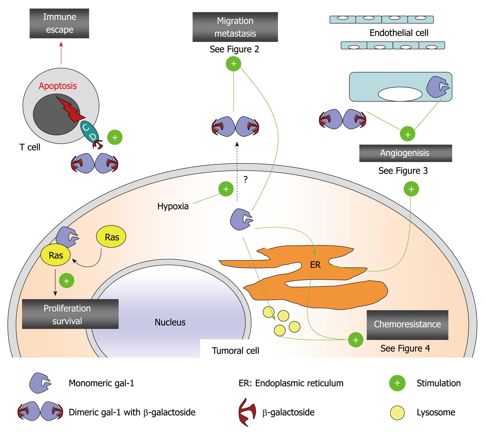
All these biological roles played by galectin-1 occur through the interactions with a myriad of extracellular and intracellular ligands and/or receptors including, but not limited to, laminin, fibronectin, vitronectin, integrins, CA-125, H-ras, CD45 and gemin-4[1-4,18,19]. Galectin-1 also plays major roles in tumor immune escape processes and in tuning the immune response. This aspect of galectin-1-related functions has been extensively reviewed[8,41-43] and is not further discussed here. Figure 1 illustrates the overall picture of galectin-1-related major roles in tumor cell biology.

Figure 1
Figure 1 Major roles of galectin-1 in tumor biology. CD: Cluster of differentiation antigen, could be CD2, CD3, CD7, CD29, CD43 and/or CD45 that are expressed at the cell surface of activated T cell and that display typical β-galactosides recognized by galectin-1.
GALECTIN-1 AND MELANOMAS

Although melanomas account for only 4% of all dermatological cancers, they are responsible for 80% of deaths from skin cancer[44,45]. In fact, the incidence of melanoma is increasing worldwide, and the prognosis for patients with high-risk or advanced metastatic melanoma remains poor despite advances in the field[44]. Patients who progress to stage IV metastatic melanoma have a median survival of < 1 year[44]. Only 14% of patients with metastatic melanoma survive for 5 years[46]. Melanomas display both intrinsic and acquired resistance to proapoptotic stimuli[47], while most chemotherapeutic agents still used to combat melanoma are proapoptotic agents[48,49].

Great hope has been placed in vaccines, but only limited successes have been observed to date[44]. Indeed, clinical trials of melanoma vaccines have yielded inconclusive data on whether a positive melanoma-specific immune response predicts treatment benefit[45]. In fact, melanoma cells are able to escape natural, endogenous or therapeutically induced immune attacks[50]. The Rabinovich group[7,8] has elegantly demonstrated that melanoma-secreted galectin-1 induces apoptosis of activated T cells through recognition of glycosylated CD3, CD7 and CD45, thereby constituting an important mechanism of tumor escape in experimental melanomas. The blockade of this inhibitory signal can allow for and potentiate effective immune responses against melanoma cells with profound implications for cancer immunotherapy. Decreasing galectin-1 expression in melanoma could be achieved through the use of anti-galectin-1 siRNA, which we have developed[51] and aim to use for vaccine therapy in glioma patients[19] as detailed below.

Melanomas are associated with marked angiogenesis[52], and hypoxia favors melanoma progression[53]. Galectin-1 is a hypoxia-inducible protein[54,55] with marked proangiogenic effects[32-35] that have been demonstrated in experimental melanomas[11]. Great hope has also been placed in antiangiogenic therapies to combat melanomas[56,57], and decreasing galectin-1 expression in melanomas would be one strategy to accomplish this.

The PubMed database already includes about 2800 publications on galectins as of September 2011 with 920 of them related to galectin-1, but only 17 publications cross-reference melanoma and galectin-1, and they often do so indirectly. In other words, little information is available about the exact roles of galectin-1 in melanoma biology. The first report was published in 1995 by van den Brûle et al[58], who have shown that galectin-1 modulates human melanoma cell adhesion to laminin. Subsequently, galectin-1 has been demonstrated to participate in the aggregation of human melanoma cells through binding to the 90K/MAC-2BP glycoprotein[59]. As mentioned above, Rabinovich and his group have demonstrated the roles of galectin-1 in immune tumor escape processes using an experimental melanoma model[7]. We have used the same model to demonstrate the protective roles of galectin-1 against chemotherapy-induced cytotoxic insults in melanoma cells[11]. Although Rondepierre et al[14] have demonstrated a direct role for galectin-1 in the biological aggressiveness of experimental melanomas, Bolander et al[12] have failed to observe any correlation between the levels of galectin-1 expression in melanoma and patient survival. Nevertheless, our recent clinical data have indicated that galectin-1 is particularly highly expressed in advanced melanoma lesions, notably in comparison to galectin-3 and -9 (submitted manuscript).

Altogether, these data that are already available in the literature strongly suggest that galectin-1 could be implicated in various biological processes linked to melanoma progression, such as tumor immune escape, tumor angiogenesis and chemoresistance. However, most of these data are from experimental models, and clinical confirmation of these findings is warranted.

GALECTIN-1 AND GLIOMAS

There are 19 publications available in the PubMed database with cross references about galectin-1 and gliomas and 17 with cross references about galectin-1 and melanomas (as of July 2011). However, much more information is available about how galectin-1 controls glioma cell biology than melanoma cell biology.

Gliomas are the most common primary brain tumors, among which glioblastoma (GBM) is the most malignant form. Malignant gliomas, especially GBMs, are characterized by the diffuse invasion of distant brain tissue by a myriad of single migrating cells with reduced levels of apoptosis and consequent resistance to the cytotoxic insults of proapoptotic drugs[60-62]. In contrast, GBM cells are less resistant to cell death induced by sustained proautophagic processes[62]. Current clinical recommendations for treating malignant glioma patients, and more specifically GBM patients, include maximum surgical resection followed by concurrent radiation and chemotherapy with temozolomide[62-64]. This clinical protocol is now the standard of care for treating GBM patients, and the overall survival rates have increased from 11% to 27% at 2 years, from 4% to 16% at 3 years, from 3% to 12% at 4 years, and from 2% to 10% at 5 years, compared to surgery and radiotherapy alone[63,64].

As detailed below, galectin-1 significantly affects glioma progression. Experimental data have already been validated, at least partly, by clinical data. By using computer-assisted microscopy, we have quantitatively characterized the levels of expression of galectins-1, 3 and 8 in 116 human astrocytic tumors of grades I-IV by immunohistochemistry. The data have indicated that the levels of galectin-1 and 3 expression significantly changes during the progression of malignancy in human astrocytic tumors, whereas galectin-8 remains unchanged[16]. We have extended our analyses to include a quantitative immunohistochemical determination of galectin-1 expression in 220 gliomas, including 151 astrocytic, 38 oligodendroglial and 31 ependymal tumors obtained from surgical resections[65]. We have also xenografted three human glioblastoma cell lines (H4, U87 and U373 models) into the brains of nude mice to characterize the in vivo galectin-1 expression pattern following subsequent invasion into the normal brain parenchyma[65]. In addition, we have characterized in vitro the role of galectin-1 in U373 tumor astrocyte migration and kinetics. Our data have revealed expression of galectin-1 in all human glioma types, with no striking differences between astrocytic, oligodendroglial and ependymal tumors[65]. The level of galectin-1 expression is correlated only with grade of astrocytic tumors[65]. Furthermore, immunopositivity of high-grade astrocytic tumors from patients with short-term survival periods is stronger than tumors from patients with long-term survival[65]. In human glioblastoma xenografts, galectin-1 is preferentially expressed in the more invasive parts of the xenografts[65]. In vitro experiments have revealed that galectin-1 stimulates the migration of U373 astrocytes[65]. Most of these data that we have produced[16,65] along with Yamaoka et al[15], have been validated by Jung et al[17].

We recently have reviewed the general roles of galectins, in particular galectin-1, in gliomas[18]. We therefore focus our attention below on the biochemical pathways in which galectin-1 is implicated when controlling various processes related to glioma progression and to a lesser extent, melanoma progression.

As mentioned earlier, we do not review in detail the marked roles exerted by galectin-1 when tuning the immune system response, and those associated with the tumor immune escape phenomenon, because all these aspects have been reviewed in depth by others[8,41-43]. Nevertheless, we highlight galectin-1 as a major target to combat gliomas when vaccine therapy is proposed for gliomas[66] and melanomas. Active specific immunotherapy based on dendritic cell vaccination is indeed considered to be a new promising concept aimed at generating an antitumoral immune response in malignant gliomas and melanomas that still have dismal prognosis despite multimodal treatments[19,66]. However, it is now widely accepted that the success of immunotherapeutic strategies to promote tumor regression will rely not only on enhancing the effector arm of the immune response, but also on the downregulation of the counteracting tolerogenic signals[19,66]. We recently reviewed why galectin-1 should be actively targeted to lower glioma-mediated immune escape during vaccine therapy[19].

GALECTIN-1 CONTROLS GLIOMA CELL MIGRATION

Although cell migration is the net result of adhesion, motility and invasion[60], galectin-1 modifies each of these three cell-migration-related processes in glioma cells.

Immunohistochemical analysis of galectin-1 expression in human U87 and U373 glioblastoma xenografts from the brains of immunodeficient mice has revealed a higher level of galectin-1 expression in invasive areas compared with non-invasive areas of the xenografts[16,65,67]. Immunodeficient mice intracranially grafted with U87 or U373 cells that constitutively express low levels of galectin-1 (by stable transfection of an expression vector containing the antisense mRNA of galectin-1) have longer survival periods than those grafted with U87 or U373 cells that express normal levels of galectin-1,[67], and galectin-1 added to the culture medium markedly increases the motility of human neoplastic astrocytes[16,65,67]. We have demonstrated that these effects are at least partly related to marked modifications in the organization of the actin cytoskeleton and increases in small GTPase RhoA expression[67]. We have also investigated stable knockdown of galectin-1 in human U87 glioblastoma cells, and observed major alterations in gene expression when we used cDNA microarray analysis. Among the 631 genes tested that are potentially involved in cancer, the expression of 86 genes was increased at least twofold[68], including ADAM-15 (disintegrin and metalloproteinase domain-containing protein 15) and microtubule-associated protein (MAP) 2, and expression of these proteins was confirmed by immunocytochemistry[68]. The major differences in the patterns of the actin stress fiber organization have also been observed[68], as previously reported for other glioma models[67], and U87 glioma cells that are stably deficient for galectin-1 expression are significantly less motile than control cells[68], as previously observed[67].

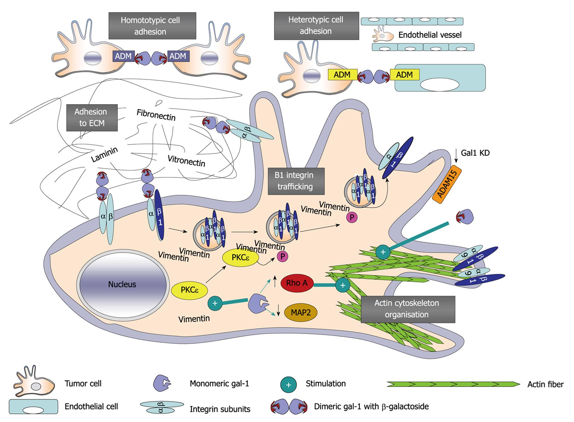
Genes whose patterns of expression markedly change during galectin-1 knockdown include α-7/β-1 and α-9/β-1 integrins[68]. These data must be analyzed in parallel with those we report above with respect to galectin-1-mediated modifications in ADAM-15 expression because the ADAM family of membrane-anchorage glycoproteins encompass a catalytically active matrix metalloproteinase domain and a disintegrin domain, and may also be involved in the proteolytic cleavage of cell-surface proteins and in integrin-mediated cell adhesion (including α-9/β-1 integrin/ADAM-15 interactions) via RGD-dependent and -independent binding[68]. Using immunofluorescence approaches, we observed that the depletion of galectin-1 through both stable knockdown and transient-targeted siRNA treatments induced an intracellular accumulation of β-1 integrin, along with a decrease in the expression of this integrin at points of adhesion on the cell membrane[69]. Galectin-1 depletion does not alter the gene expression level of β-1 integrin[69]. Transient galectin-1 depletion induces perinuclear accumulation of protein kinase C (PKC)ε and intermediate filament vimentin, both of which have been shown to mediate integrin recycling in motile cells. These data emphasize the involvement of galectin-1 in the PKCε/vimentin-controlled trafficking of β-1 integrin in glioma cells. Figure 2 illustrates the gross picture of galectin-1-related major roles in glioma cell migration.

Figure 2
Figure 2 Processes and pathways mediated by galectin-1 in cancer cell migration. ADM: Adhesion molecules; ADAM-15: Disintegrin and metalloproteinase domain-containing protein 15; ECM: Extracellular matrix; MAP2: Microtubule-associated protein 2; P: Phosphorylation; PKCε: Protein kinase C ε; RhoA: Ras homolog protein, member A GTPase.
GALECTIN-1 CONTROLS NEOANGIOGENESIS IN GLIOMAS AND MELANOMAS

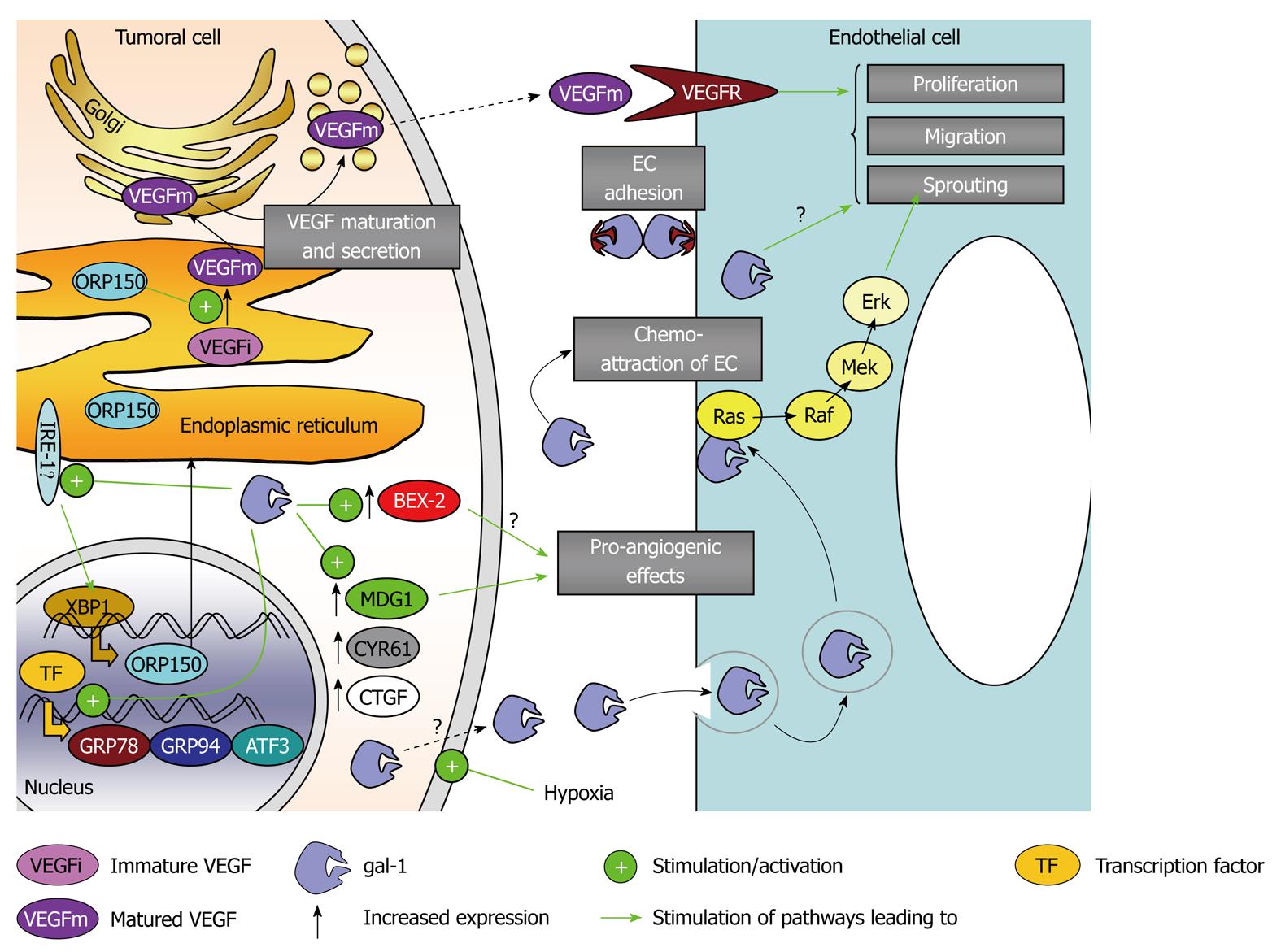
As mentioned earlier, galectin-1 is a hypoxia-regulated protein[54,55] that has been shown to have major roles in angiogenesis[32,35] of gliomas[33] and melanomas[11]. We first highlighted that galectin-1 depleted melanoma tumors in vivo display lower angiogenesis levels in close association with marked necrotic processes[11]. We have recently begun to decipher the molecular and biochemical pathways through which galectin-1 controls angiogenesis. In gliomas, we have demonstrated that galectin-1 signals through the IRE-1α (endoplasmic reticulum transmembrane kinase/ribonuclease inositol-requiring 1α), which regulates the expression of oxygen-regulated protein 150 (ORP150) that in turn controls vascular endothelial growth factor (VEGF) maturation[33]. Thus, galectin-1 controls glioma angiogenesis through ORP150-mediated VEGF maturation[33]. Similarly, galectin-1 depletion is associated with decreased ORP150 expression level in melanoma cells (unpublished data). Galectin-1 also modulates the expression of several other hypoxia-related genes (e.g. CTGF, ATF3, PPP1R15A, HSPA5, TRA1 and CYR61) in the Hs683 glioma model, which are known to display various roles in angiogenesis, which we have demonstrated in galectin-1-dependent angiogenesis in glioma[33].

We have observed a marked decrease in the expression of the brain-expressed X-linked gene, BEX2, with decreased galectin-1 expression in glioma cells through targeted anti-galectin-1 siRNA[34]. We have thus focused on BEX2, and observed that decreasing BEX2 expression in human Hs683 glioma cells increased the survival of Hs683 orthotopic xenograft-bearing immunodeficient mice, whose tumors displayed decreased angiogenic levels[34]. Furthermore, this decrease in BEX2 expression impaired vasculogenic mimicry channel formation in vitro, as observed when depleting galectin-1 in both glioma and melanoma cells. Thus, BEX2 is a second target, in addition to ORP150, through which galectin-1 controls angiogenesis in gliomas.

BEX2 also modulates glioma cell migration at both adhesion and invasion levels through the modification of several genes previously reported to play a role in cancer cell migration, including MAP2, plexin C1, SWAP70, and β-6 integrin[34].

Galectin-1 controls key angiogenic factors/ pathways in tumoral cells. In addition, galectin-1 has been shown to regulate directly the biological properties of endothelial cells, such as proliferation, activation and in vitro tubular network formation[32,35]. We have observed earlier that galectin-3 also participates in glioma angiogenesis[70].

GALECTIN-1 AND ITS MODULATING ROLES IN CHEMOTHERAPY AND RADIOTHERAPY IN THE SPECIFIC CONTEXT OF MELANOMAS AND GLIOMAS

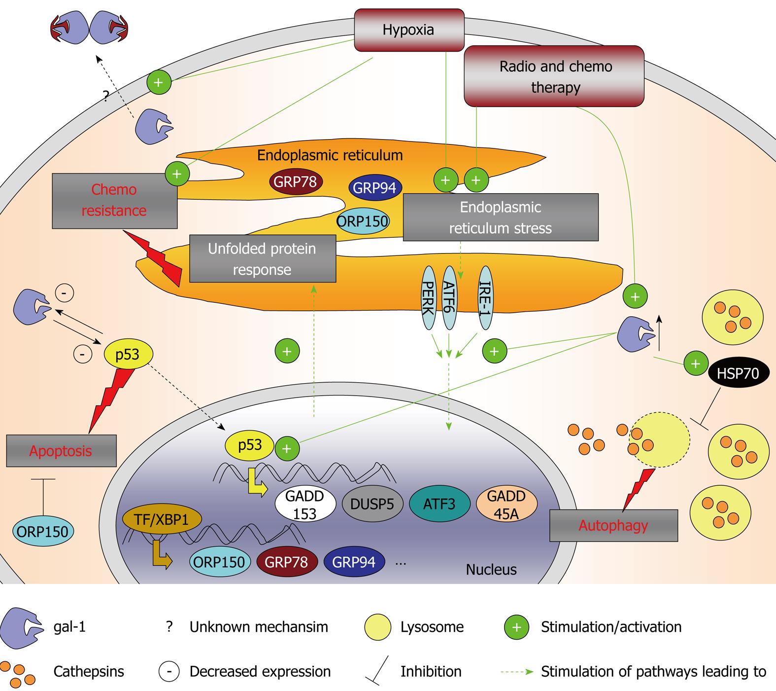
Intratumoral hypoxia causes genetic changes in cancers that produce a microenvironment that selects for cells with a more aggressive phenotype[71]. Hypoxia can initiate cell demise by apoptosis/necrosis but can also prevent cell death by provoking adaptive responses that facilitate cell proliferation or angiogenesis, thus contributing to tumor malignant progression[71], and in particular, gliomas[72] and melanomas[53]. Hypoxia is also known to modulate the UPR, a coordinated program that promotes cell survival under conditions of endoplasmic reticulum (ER) stress, which is known to contribute to tumor malignant progression and drug resistance of solid tumors[73]. Considering that galectin-1 is a hypoxia-regulated protein[54,55], we have investigated whether it can interfere with ER stress responses and chemosensitivity. We have examined whether decreasing galectin-1 expression (by means of a siRNA approach) in human Hs683 GBM cells and B16F10 melanoma cells could increase their sensitivity to pro-autophagic or proapoptotic drugs. These data have revealed that temozolomide, the standard treatment for glioma patients, increases galectin-1 expression in Hs683 cells both in vitro[36] and in vivo[33]. In contrast, reducing galectin-1 expression in these Hs683 glioma cells using siRNA increases the antitumor effects of various chemotherapeutic agents, in particular temozolomide, both in vitro and in vivo[33,36], which is a feature that we also have observed in experimental melanomas[11]. This decrease in galectin-1 expression in Hs683 glioma cells does not induce apoptotic or autophagic features, but is found to modulate p53 transcriptional activity and decrease p53-targeted gene expression including DDIT3/GADD153/CHOP, DUSP5, ATF3 and GADD45A[36]. Puchades et al[74] have demonstrated that galectin-1 expression is negatively regulated by transfection with TP53 in glioma cells. We have further observed that the decrease in galectin-1 expression in glioma cells also impairs the expression levels of seven other genes implicated in chemoresistance: ORP150, HERP, GRP78/Bip, TRA1, BNIP3L, GADD45B and CYR61; some of which are located in the ER and whose expression is also known to be modified by hypoxia[36]. In the case of the B16F10 mouse melanoma model, decreasing galectin-1 expression in vitro by means of an anti-galectin-1 siRNA approach does not modify their sensitivity to apoptosis or autophagy[11]. However, it does induce heat-shock-protein-70-mediated lysosomal membrane permeabilization, a process associated with cathepsin B release into the cytosol, which in turn is believed to sensitize the cells to the pro-autophagic effects of temozolomide when grafted in vivo[11].

Galectin-1 expression is also upregulated by ionizing irradiation in glioma cell lines[37]. Therefore galectin-1 appears to be induced in cases of various cellular stress stimuli and could promote cell survival through the various mechanisms described above and illustrated in Figure 3.

Figure 3
Figure 3 Galectin-1-mediated control of angiogenesis. ATF3: Activating transcription factor 3; BEX2: Brain-expressed X-linked 2; CTGF: Connective tissue growth factor; CYR61: Cysteine-rich angiogenic inducer 61; Erk: Extracellular signal-regulated kinase; GRP78 and GRP94: Glucose-regulated protein 78 and 94; IRE1-α: Endoplasmic reticulum to nucleus signaling 1 α; MDG1: Microvascular endothelial differentiation gene 1; Mek: Mitogen-activated protein kinase kinase; ORP150: Oxygen-related protein 150; Raf: Homolog to murine v-raf leukemia oncogene serine/threonine kinase; Ras: Rat sarcoma virus oncogene protein; TF: Transcription factor; VEGF: Vascular endothelial growth factor; VEGFR: VEGF receptor; XBP1: X-box-binding protein 1. EC: Endothelial cell.
CONCLUSION

Gliomas and melanomas are associated with dismal prognosis because of their marked intrinsic resistance to proapoptotic stimuli such as conventional chemotherapy and radiotherapy, as well as their capability to escape immune cell attacks. In addition, gliomas and melanomas display pronounced neoangiogenesis. Galectin-1 is a hypoxia-sensitive protein that is abundantly secreted by glioma and melanoma cells, displays marked proangiogenic effects, and provides immunotolerogenic environments to melanoma and glioma cells through the killing of activated T cells that attack these tumor cells. Galectin-1 also protects glioma and melanoma cells against cytotoxic insults (chemotherapy and radiotherapy) through a direct role in the UPR. Altogether, these facts clearly point to galectin-1 as an important target in gliomas and melanomas, to weaken the defenses of these two types of cancers against radiotherapy, chemotherapy and immunotherapy/vaccine therapy, and to reinforce antiangiogenic therapies Figure 4.

Figure 4
Figure 4 How galectin-1 is involved in chemoresistance of cancer cells. ATF3 and ATF6: Activating transcription factor 3 and 6; DUSP5: Dual specificity protein phosphatase 5; GADD153 and GADD45A: Growth arrest and DNA damage inducible protein 153 and 45 α; GRP78 and GRP94: Glucose-regulated protein 78 and 94; HSP70: Heat shock protein 70; IRE1-α: Endoplasmic reticulum to nucleus signaling 1 α; ORP150: Oxygen-related protein 150; PERK: Protein kinase RNA-like endoplasmic reticulum kinase; TF: Transcription factor; XBP1: X-box-binding protein 1.
Footnotes

Peer reviewers: Ming Zhang, PhD, Associate Professor, Department of Molecular Pharmacology and Biological Chemistry, Feinberg School of Medicine Northwestern University, 303 E. Superior St., Chicago, IL 60611, United States; Shile Huang, PhD, Associate Professor, Department of Biochemistry and Molecular Biology, Louisiana State University Health Sciences Center, Shreveport, LA 71130-3932, United States; Tatjana Abaffy, Dr, Molecular and Cellular Pharmacology, University of Miami, Miller School of Medicine, 1600 NW 10 Ave, Miami, FL 33136, United States

S- Editor Cheng JX L- Editor Kerr C E- Editor Zheng XM

References
1.  Barondes SH, Cooper DN, Gitt MA, Leffler H. Galectins. Structure and function of a large family of animal lectins. J Biol Chem. 1994;269:20807-20810.  [PubMed]  [DOI]  [Cited in This Article: ]
2.  Danguy A, Camby I, Kiss R. Galectins and cancer. Biochim Biophys Acta. 2002;1572:285-293.  [PubMed]  [DOI]  [Cited in This Article: ]
3.  Liu FT, Rabinovich GA. Galectins as modulators of tumour progression. Nat Rev Cancer. 2005;5:29-41.  [PubMed]  [DOI]  [Cited in This Article: ]  [Cited by in Crossref: 1074]  [Cited by in F6Publishing: 1078]  [Article Influence: 56.7]  [Reference Citation Analysis (0)]
4.  Camby I, Le Mercier M, Lefranc F, Kiss R. Galectin-1: a small protein with major functions. Glycobiology. 2006;16:137R-157R.  [PubMed]  [DOI]  [Cited in This Article: ]  [Cited by in Crossref: 595]  [Cited by in F6Publishing: 653]  [Article Influence: 36.3]  [Reference Citation Analysis (0)]
5.  Houzelstein D, Gonçalves IR, Fadden AJ, Sidhu SS, Cooper DN, Drickamer K, Leffler H, Poirier F. Phylogenetic analysis of the vertebrate galectin family. Mol Biol Evol. 2004;21:1177-1187.  [PubMed]  [DOI]  [Cited in This Article: ]  [Cited by in Crossref: 196]  [Cited by in F6Publishing: 193]  [Article Influence: 9.7]  [Reference Citation Analysis (0)]
6.  Perillo NL, Pace KE, Seilhamer JJ, Baum LG. Apoptosis of T cells mediated by galectin-1. Nature. 1995;378:736-739.  [PubMed]  [DOI]  [Cited in This Article: ]  [Cited by in Crossref: 777]  [Cited by in F6Publishing: 779]  [Article Influence: 26.9]  [Reference Citation Analysis (0)]
7.  Rubinstein N, Alvarez M, Zwirner NW, Toscano MA, Ilarregui JM, Bravo A, Mordoh J, Fainboim L, Podhajcer OL, Rabinovich GA. Targeted inhibition of galectin-1 gene expression in tumor cells results in heightened T cell-mediated rejection; A potential mechanism of tumor-immune privilege. Cancer Cell. 2004;5:241-251.  [PubMed]  [DOI]  [Cited in This Article: ]  [Cited by in Crossref: 403]  [Cited by in F6Publishing: 397]  [Article Influence: 19.9]  [Reference Citation Analysis (0)]
8.  Rabinovich GA, Ilarregui JM. Conveying glycan information into T-cell homeostatic programs: a challenging role for galectin-1 in inflammatory and tumor microenvironments. Immunol Rev. 2009;230:144-159.  [PubMed]  [DOI]  [Cited in This Article: ]  [Cited by in Crossref: 66]  [Cited by in F6Publishing: 66]  [Article Influence: 4.4]  [Reference Citation Analysis (0)]
9.  Ilarregui JM, Croci DO, Bianco GA, Toscano MA, Salatino M, Vermeulen ME, Geffner JR, Rabinovich GA. Tolerogenic signals delivered by dendritic cells to T cells through a galectin-1-driven immunoregulatory circuit involving interleukin 27 and interleukin 10. Nat Immunol. 2009;10:981-991.  [PubMed]  [DOI]  [Cited in This Article: ]  [Cited by in Crossref: 325]  [Cited by in F6Publishing: 339]  [Article Influence: 22.6]  [Reference Citation Analysis (0)]
10.  Rabinovich GA. Galectin-1 as a potential cancer target. Br J Cancer. 2005;92:1188-1192.  [PubMed]  [DOI]  [Cited in This Article: ]  [Cited by in Crossref: 169]  [Cited by in F6Publishing: 176]  [Article Influence: 9.3]  [Reference Citation Analysis (0)]
11.  Mathieu V, Le Mercier M, De Neve N, Sauvage S, Gras T, Roland I, Lefranc F, Kiss R. Galectin-1 knockdown increases sensitivity to temozolomide in a B16F10 mouse metastatic melanoma model. J Invest Dermatol. 2007;127:2399-2410.  [PubMed]  [DOI]  [Cited in This Article: ]  [Cited by in Crossref: 73]  [Cited by in F6Publishing: 74]  [Article Influence: 4.4]  [Reference Citation Analysis (0)]
12.  Bolander A, Agnarsdóttir M, Strömberg S, Ponten F, Hesselius P, Uhlen M, Bergqvist M. The protein expression of TRP-1 and galectin-1 in cutaneous malignant melanomas. Cancer Genomics Proteomics. 2008;5:293-300.  [PubMed]  [DOI]  [Cited in This Article: ]
13.  Stessl M, Marchetti-Deschmann M, Winkler J, Lachmann B, Allmaier G, Noe CR. A proteomic study reveals unspecific apoptosis induction and reduction of glycolytic enzymes by the phosphorothioate antisense oligonucleotide oblimersen in human melanoma cells. J Proteomics. 2009;72:1019-1030.  [PubMed]  [DOI]  [Cited in This Article: ]  [Cited by in Crossref: 24]  [Cited by in F6Publishing: 24]  [Article Influence: 1.6]  [Reference Citation Analysis (0)]
14.  Rondepierre F, Bouchon B, Bonnet M, Moins N, Chezal JM, D'Incan M, Degoul F. B16 melanoma secretomes and in vitro invasiveness: syntenin as an invasion modulator. Melanoma Res. 2010;20:77-84.  [PubMed]  [DOI]  [Cited in This Article: ]  [Cited by in Crossref: 11]  [Cited by in F6Publishing: 13]  [Article Influence: 0.9]  [Reference Citation Analysis (0)]
15.  Yamaoka K, Mishima K, Nagashima Y, Asai A, Sanai Y, Kirino T. Expression of galectin-1 mRNA correlates with the malignant potential of human gliomas and expression of antisense galectin-1 inhibits the growth of 9 glioma cells. J Neurosci Res. 2000;59:722-730.  [PubMed]  [DOI]  [Cited in This Article: ]
16.  Camby I, Belot N, Rorive S, Lefranc F, Maurage CA, Lahm H, Kaltner H, Hadari Y, Ruchoux MM, Brotchi J. Galectins are differentially expressed in supratentorial pilocytic astrocytomas, astrocytomas, anaplastic astrocytomas and glioblastomas, and significantly modulate tumor astrocyte migration. Brain Pathol. 2001;11:12-26.  [PubMed]  [DOI]  [Cited in This Article: ]  [Cited by in Crossref: 118]  [Cited by in F6Publishing: 130]  [Article Influence: 7.2]  [Reference Citation Analysis (0)]
17.  Jung TY, Jung S, Ryu HH, Jeong YI, Jin YH, Jin SG, Kim IY, Kang SS, Kim HS. Role of galectin-1 in migration and invasion of human glioblastoma multiforme cell lines. J Neurosurg. 2008;109:273-284.  [PubMed]  [DOI]  [Cited in This Article: ]  [Cited by in Crossref: 43]  [Cited by in F6Publishing: 44]  [Article Influence: 2.8]  [Reference Citation Analysis (0)]
18.  Le Mercier M, Fortin S, Mathieu V, Kiss R, Lefranc F. Galectins and gliomas. Brain Pathol. 2010;20:17-27.  [PubMed]  [DOI]  [Cited in This Article: ]  [Cited by in Crossref: 79]  [Cited by in F6Publishing: 82]  [Article Influence: 5.5]  [Reference Citation Analysis (0)]
19.  Verschuere T, De Vleeschouwer S, Lefranc F, Kiss R, Van Gool SW. Galectin-1 and immunotherapy for brain cancer. Expert Rev Neurother. 2011;11:533-543.  [PubMed]  [DOI]  [Cited in This Article: ]  [Cited by in Crossref: 20]  [Cited by in F6Publishing: 22]  [Article Influence: 1.7]  [Reference Citation Analysis (0)]
20.  Strik HM, Kolodziej M, Oertel W, Bäsecke J. Glycobiology in Malignant Gliomas: Expression and Functions of Galectins and Possible Therapeutic Options. Curr Pharm Biotechnol. 2011;Epub ahead of print.  [PubMed]  [DOI]  [Cited in This Article: ]
21.  Seelenmeyer C, Wegehingel S, Tews I, Künzler M, Aebi M, Nickel W. Cell surface counter receptors are essential components of the unconventional export machinery of galectin-1. J Cell Biol. 2005;171:373-381.  [PubMed]  [DOI]  [Cited in This Article: ]  [Cited by in Crossref: 87]  [Cited by in F6Publishing: 84]  [Article Influence: 4.7]  [Reference Citation Analysis (0)]
22.  Seelenmeyer C, Stegmayer C, Nickel W. Unconventional secretion of fibroblast growth factor 2 and galectin-1 does not require shedding of plasma membrane-derived vesicles. FEBS Lett. 2008;582:1362-1368.  [PubMed]  [DOI]  [Cited in This Article: ]  [Cited by in Crossref: 48]  [Cited by in F6Publishing: 39]  [Article Influence: 2.4]  [Reference Citation Analysis (0)]
23.  Elad-Sfadia G, Haklai R, Ballan E, Gabius HJ, Kloog Y. Galectin-1 augments Ras activation and diverts Ras signals to Raf-1 at the expense of phosphoinositide 3-kinase. J Biol Chem. 2002;277:37169-37175.  [PubMed]  [DOI]  [Cited in This Article: ]  [Cited by in Crossref: 166]  [Cited by in F6Publishing: 176]  [Article Influence: 8.0]  [Reference Citation Analysis (0)]
24.  Rotblat B, Niv H, André S, Kaltner H, Gabius HJ, Kloog Y. Galectin-1(L11A) predicted from a computed galectin-1 farnesyl-binding pocket selectively inhibits Ras-GTP. Cancer Res. 2004;64:3112-3118.  [PubMed]  [DOI]  [Cited in This Article: ]  [Cited by in Crossref: 121]  [Cited by in F6Publishing: 126]  [Article Influence: 6.3]  [Reference Citation Analysis (0)]
25.  Scott K, Weinberg C. Galectin-1: a bifunctional regulator of cellular proliferation. Glycoconj J. 2004;19:467-477.  [PubMed]  [DOI]  [Cited in This Article: ]  [Cited by in F6Publishing: 1]  [Reference Citation Analysis (0)]
26.  He J, Baum LG. Galectin interactions with extracellular matrix and effects on cellular function. Methods Enzymol. 2006;417:247-256.  [PubMed]  [DOI]  [Cited in This Article: ]  [Cited by in Crossref: 63]  [Cited by in F6Publishing: 66]  [Article Influence: 3.7]  [Reference Citation Analysis (0)]
27.  Hsu DK, Liu FT. Regulation of cellular homeostasis by galectins. Glycoconj J. 2004;19:507-515.  [PubMed]  [DOI]  [Cited in This Article: ]  [Cited by in F6Publishing: 1]  [Reference Citation Analysis (0)]
28.  Yang RY, Liu FT. Galectins in cell growth and apoptosis. Cell Mol Life Sci. 2003;60:267-276.  [PubMed]  [DOI]  [Cited in This Article: ]  [Cited by in Crossref: 146]  [Cited by in F6Publishing: 150]  [Article Influence: 7.1]  [Reference Citation Analysis (0)]
29.  Delbrouck C, Doyen I, Belot N, Decaestecker C, Ghanooni R, de Lavareille A, Kaltner H, Choufani G, Danguy A, Vandenhoven G. Galectin-1 is overexpressed in nasal polyps under budesonide and inhibits eosinophil migration. Lab Invest. 2002;82:147-158.  [PubMed]  [DOI]  [Cited in This Article: ]
30.  Barrow H, Rhodes JM, Yu LG. The role of galectins in colorectal cancer progression. Int J Cancer. 2011;129:1-8.  [PubMed]  [DOI]  [Cited in This Article: ]  [Cited by in Crossref: 74]  [Cited by in F6Publishing: 79]  [Article Influence: 6.1]  [Reference Citation Analysis (0)]
31.  Fulcher JA, Hashimi ST, Levroney EL, Pang M, Gurney KB, Baum LG, Lee B. Galectin-1-matured human monocyte-derived dendritic cells have enhanced migration through extracellular matrix. J Immunol. 2006;177:216-226.  [PubMed]  [DOI]  [Cited in This Article: ]
32.  Thijssen VL, Postel R, Brandwijk RJ, Dings RP, Nesmelova I, Satijn S, Verhofstad N, Nakabeppu Y, Baum LG, Bakkers J. Galectin-1 is essential in tumor angiogenesis and is a target for antiangiogenesis therapy. Proc Natl Acad Sci USA. 2006;103:15975-15980.  [PubMed]  [DOI]  [Cited in This Article: ]  [Cited by in Crossref: 351]  [Cited by in F6Publishing: 358]  [Article Influence: 19.9]  [Reference Citation Analysis (0)]
33.  Le Mercier M, Mathieu V, Haibe-Kains B, Bontempi G, Mijatovic T, Decaestecker C, Kiss R, Lefranc F. Knocking down galectin 1 in human hs683 glioblastoma cells impairs both angiogenesis and endoplasmic reticulum stress responses. J Neuropathol Exp Neurol. 2008;67:456-469.  [PubMed]  [DOI]  [Cited in This Article: ]
34.  Le Mercier M, Fortin S, Mathieu V, Roland I, Spiegl-Kreinecker S, Haibe-Kains B, Bontempi G, Decaestecker C, Berger W, Lefranc F. Galectin 1 proangiogenic and promigratory effects in the Hs683 oligodendroglioma model are partly mediated through the control of BEX2 expression. Neoplasia. 2009;11:485-496.  [PubMed]  [DOI]  [Cited in This Article: ]
35.  Thijssen VL, Barkan B, Shoji H, Aries IM, Mathieu V, Deltour L, Hackeng TM, Kiss R, Kloog Y, Poirier F. Tumor cells secrete galectin-1 to enhance endothelial cell activity. Cancer Res. 2010;70:6216-6224.  [PubMed]  [DOI]  [Cited in This Article: ]  [Cited by in Crossref: 173]  [Cited by in F6Publishing: 184]  [Article Influence: 13.1]  [Reference Citation Analysis (0)]
36.  Le Mercier M, Lefranc F, Mijatovic T, Debeir O, Haibe-Kains B, Bontempi G, Decaestecker C, Kiss R, Mathieu V. Evidence of galectin-1 involvement in glioma chemoresistance. Toxicol Appl Pharmacol. 2008;229:172-183.  [PubMed]  [DOI]  [Cited in This Article: ]  [Cited by in Crossref: 76]  [Cited by in F6Publishing: 64]  [Article Influence: 4.0]  [Reference Citation Analysis (0)]
37.  Strik HM, Schmidt K, Lingor P, Tönges L, Kugler W, Nitsche M, Rabinovich GA, Bähr M. Galectin-1 expression in human glioma cells: modulation by ionizing radiation and effects on tumor cell proliferation and migration. Oncol Rep. 2007;18:483-488.  [PubMed]  [DOI]  [Cited in This Article: ]
38.  Wang W, Park JW, Wang JL, Patterson RJ. Immunoprecipitation of spliceosomal RNAs by antisera to galectin-1 and galectin-3. Nucleic Acids Res. 2006;34:5166-5174.  [PubMed]  [DOI]  [Cited in This Article: ]  [Cited by in Crossref: 31]  [Cited by in F6Publishing: 31]  [Article Influence: 1.7]  [Reference Citation Analysis (0)]
39.  Voss PG, Gray RM, Dickey SW, Wang W, Park JW, Kasai K, Hirabayashi J, Patterson RJ, Wang JL. Dissociation of the carbohydrate-binding and splicing activities of galectin-1. Arch Biochem Biophys. 2008;478:18-25.  [PubMed]  [DOI]  [Cited in This Article: ]  [Cited by in Crossref: 19]  [Cited by in F6Publishing: 20]  [Article Influence: 1.3]  [Reference Citation Analysis (0)]
40.  Haudek KC, Patterson RJ, Wang JL. SR proteins and galectins: what's in a name? Glycobiology. 2010;20:1199-1207.  [PubMed]  [DOI]  [Cited in This Article: ]  [Cited by in Crossref: 36]  [Cited by in F6Publishing: 36]  [Article Influence: 2.6]  [Reference Citation Analysis (0)]
41.  Rabinovich GA, Liu FT, Hirashima M, Anderson A. An emerging role for galectins in tuning the immune response: lessons from experimental models of inflammatory disease, autoimmunity and cancer. Scand J Immunol. 2007;66:143-158.  [PubMed]  [DOI]  [Cited in This Article: ]  [Cited by in Crossref: 185]  [Cited by in F6Publishing: 190]  [Article Influence: 11.2]  [Reference Citation Analysis (0)]
42.  Dhirapong A, Lleo A, Leung P, Gershwin ME, Liu FT. The immunological potential of galectin-1 and -3. Autoimmun Rev. 2009;8:360-363.  [PubMed]  [DOI]  [Cited in This Article: ]  [Cited by in Crossref: 76]  [Cited by in F6Publishing: 83]  [Article Influence: 5.2]  [Reference Citation Analysis (0)]
43.  Cooper D, Ilarregui JM, Pesoa SA, Croci DO, Perretti M, Rabinovich GA. Multiple functional targets of the immunoregulatory activity of galectin-1: Control of immune cell trafficking, dendritic cell physiology, and T-cell fate. Methods Enzymol. 2010;480:199-244.  [PubMed]  [DOI]  [Cited in This Article: ]  [Cited by in Crossref: 33]  [Cited by in F6Publishing: 35]  [Article Influence: 2.5]  [Reference Citation Analysis (0)]
44.  Garbe C, Eigentler TK, Keilholz U, Hauschild A, Kirkwood JM. Systematic review of medical treatment in melanoma: current status and future prospects. Oncologist. 2011;16:5-24.  [PubMed]  [DOI]  [Cited in This Article: ]  [Cited by in Crossref: 406]  [Cited by in F6Publishing: 388]  [Article Influence: 29.8]  [Reference Citation Analysis (0)]
45.  Chi M, Dudek AZ. Vaccine therapy for metastatic melanoma: systematic review and meta-analysis of clinical trials. Melanoma Res. 2011;21:165-174.  [PubMed]  [DOI]  [Cited in This Article: ]  [Cited by in Crossref: 31]  [Cited by in F6Publishing: 32]  [Article Influence: 2.5]  [Reference Citation Analysis (0)]
46.  Miller AJ, Mihm MC. Melanoma. N Engl J Med. 2006;355:51-65.  [PubMed]  [DOI]  [Cited in This Article: ]  [Cited by in Crossref: 1083]  [Cited by in F6Publishing: 1035]  [Article Influence: 57.5]  [Reference Citation Analysis (0)]
47.  Soengas MS, Lowe SW. Apoptosis and melanoma chemoresistance. Oncogene. 2003;22:3138-3151.  [PubMed]  [DOI]  [Cited in This Article: ]  [Cited by in Crossref: 605]  [Cited by in F6Publishing: 606]  [Article Influence: 28.9]  [Reference Citation Analysis (0)]
48.  Hersey P. Apoptosis and melanoma: how new insights are effecting the development of new therapies for melanoma. Curr Opin Oncol. 2006;18:189-196.  [PubMed]  [DOI]  [Cited in This Article: ]  [Cited by in Crossref: 37]  [Cited by in F6Publishing: 38]  [Article Influence: 2.1]  [Reference Citation Analysis (0)]
49.  La Porta CA. Mechanism of drug sensitivity and resistance in melanoma. Curr Cancer Drug Targets. 2009;9:391-397.  [PubMed]  [DOI]  [Cited in This Article: ]  [Cited by in Crossref: 47]  [Cited by in F6Publishing: 51]  [Article Influence: 3.4]  [Reference Citation Analysis (0)]
50.  Gajewski TF. Failure at the effector phase: immune barriers at the level of the melanoma tumor microenvironment. Clin Cancer Res. 2007;13:5256-5261.  [PubMed]  [DOI]  [Cited in This Article: ]  [Cited by in Crossref: 170]  [Cited by in F6Publishing: 177]  [Article Influence: 10.4]  [Reference Citation Analysis (0)]
51.  Camby I, Henriet P, Lefranc F, Courtoy P, Kiss R. Use of galectin-1-targeted RNAi-based approach for the treatment of glioma. US patent. 2006;.  [PubMed]  [DOI]  [Cited in This Article: ]
52.  Helfrich I, Schadendorf D. Blood vessel maturation, vascular phenotype and angiogenic potential in malignant melanoma: one step forward for overcoming anti-angiogenic drug resistance? Mol Oncol. 2011;5:137-149.  [PubMed]  [DOI]  [Cited in This Article: ]  [Cited by in Crossref: 37]  [Cited by in F6Publishing: 39]  [Article Influence: 3.0]  [Reference Citation Analysis (0)]
53.  Bedogni B, Powell MB. Hypoxia, melanocytes and melanoma - survival and tumor development in the permissive microenvironment of the skin. Pigment Cell Melanoma Res. 2009;22:166-174.  [PubMed]  [DOI]  [Cited in This Article: ]  [Cited by in Crossref: 84]  [Cited by in F6Publishing: 86]  [Article Influence: 5.7]  [Reference Citation Analysis (0)]
54.  Le QT, Shi G, Cao H, Nelson DW, Wang Y, Chen EY, Zhao S, Kong C, Richardson D, O'Byrne KJ. Galectin-1: a link between tumor hypoxia and tumor immune privilege. J Clin Oncol. 2005;23:8932-8941.  [PubMed]  [DOI]  [Cited in This Article: ]  [Cited by in Crossref: 187]  [Cited by in F6Publishing: 186]  [Article Influence: 9.8]  [Reference Citation Analysis (0)]
55.  Zhao XY, Chen TT, Xia L, Guo M, Xu Y, Yue F, Jiang Y, Chen GQ, Zhao KW. Hypoxia inducible factor-1 mediates expression of galectin-1: the potential role in migration/invasion of colorectal cancer cells. Carcinogenesis. 2010;31:1367-1375.  [PubMed]  [DOI]  [Cited in This Article: ]  [Cited by in Crossref: 101]  [Cited by in F6Publishing: 106]  [Article Influence: 7.6]  [Reference Citation Analysis (0)]
56.  Marneros AG. Tumor angiogenesis in melanoma. Hematol Oncol Clin North Am. 2009;23:431-46, vii-viii.  [PubMed]  [DOI]  [Cited in This Article: ]  [Cited by in Crossref: 22]  [Cited by in F6Publishing: 17]  [Article Influence: 1.1]  [Reference Citation Analysis (0)]
57.  Mansfield AS, Markovic SN. Novel therapeutics for the treatment of metastatic melanoma. Future Oncol. 2009;5:543-557.  [PubMed]  [DOI]  [Cited in This Article: ]  [Cited by in Crossref: 15]  [Cited by in F6Publishing: 15]  [Article Influence: 1.0]  [Reference Citation Analysis (0)]
58.  van den Brûle FA, Buicu C, Baldet M, Sobel ME, Cooper DN, Marschal P, Castronovo V. Galectin-1 modulates human melanoma cell adhesion to laminin. Biochem Biophys Res Commun. 1995;209:760-767.  [PubMed]  [DOI]  [Cited in This Article: ]
59.  Tinari N, Kuwabara I, Huflejt ME, Shen PF, Iacobelli S, Liu FT. Glycoprotein 90K/MAC-2BP interacts with galectin-1 and mediates galectin-1-induced cell aggregation. Int J Cancer. 2001;91:167-172.  [PubMed]  [DOI]  [Cited in This Article: ]
60.  Lefranc F, Brotchi J, Kiss R. Possible future issues in the treatment of glioblastomas: special emphasis on cell migration and the resistance of migrating glioblastoma cells to apoptosis. J Clin Oncol. 2005;23:2411-2422.  [PubMed]  [DOI]  [Cited in This Article: ]  [Cited by in Crossref: 418]  [Cited by in F6Publishing: 411]  [Article Influence: 21.6]  [Reference Citation Analysis (0)]
61.  Lefranc F, Sadeghi N, Camby I, Metens T, Dewitte O, Kiss R. Present and potential future issues in glioblastoma treatment. Expert Rev Anticancer Ther. 2006;6:719-732.  [PubMed]  [DOI]  [Cited in This Article: ]
62.  Lefranc F, Facchini V, Kiss R. Proautophagic drugs: a novel means to combat apoptosis-resistant cancers, with a special emphasis on glioblastomas. Oncologist. 2007;12:1395-1403.  [PubMed]  [DOI]  [Cited in This Article: ]
63.  Stupp R, Mason WP, van den Bent MJ, Weller M, Fisher B, Taphoorn MJ, Belanger K, Brandes AA, Marosi C, Bogdahn U. Radiotherapy plus concomitant and adjuvant temozolomide for glioblastoma. N Engl J Med. 2005;352:987-996.  [PubMed]  [DOI]  [Cited in This Article: ]  [Cited by in Crossref: 14033]  [Cited by in F6Publishing: 14602]  [Article Influence: 768.5]  [Reference Citation Analysis (0)]
64.  Stupp R, Hegi ME, Mason WP, van den Bent MJ, Taphoorn MJ, Janzer RC, Ludwin SK, Allgeier A, Fisher B, Belanger K. Effects of radiotherapy with concomitant and adjuvant temozolomide versus radiotherapy alone on survival in glioblastoma in a randomised phase III study: 5-year analysis of the EORTC-NCIC trial. Lancet Oncol. 2009;10:459-466.  [PubMed]  [DOI]  [Cited in This Article: ]  [Cited by in Crossref: 4968]  [Cited by in F6Publishing: 5510]  [Article Influence: 367.3]  [Reference Citation Analysis (1)]
65.  Rorive S, Belot N, Decaestecker C, Lefranc F, Gordower L, Micik S, Maurage CA, Kaltner H, Ruchoux MM, Danguy A. Galectin-1 is highly expressed in human gliomas with relevance for modulation of invasion of tumor astrocytes into the brain parenchyma. Glia. 2001;33:241-255.  [PubMed]  [DOI]  [Cited in This Article: ]
66.  Van Gool S, Maes W, Ardon H, Verschuere T, Van Cauter S, De Vleeschouwer S. Dendritic cell therapy of high-grade gliomas. Brain Pathol. 2009;19:694-712.  [PubMed]  [DOI]  [Cited in This Article: ]  [Cited by in Crossref: 65]  [Cited by in F6Publishing: 75]  [Article Influence: 5.0]  [Reference Citation Analysis (0)]
67.  Camby I, Belot N, Lefranc F, Sadeghi N, de Launoit Y, Kaltner H, Musette S, Darro F, Danguy A, Salmon I. Galectin-1 modulates human glioblastoma cell migration into the brain through modifications to the actin cytoskeleton and levels of expression of small GTPases. J Neuropathol Exp Neurol. 2002;61:585-596.  [PubMed]  [DOI]  [Cited in This Article: ]
68.  Camby I, Decaestecker C, Lefranc F, Kaltner H, Gabius HJ, Kiss R. Galectin-1 knocking down in human U87 glioblastoma cells alters their gene expression pattern. Biochem Biophys Res Commun. 2005;335:27-35.  [PubMed]  [DOI]  [Cited in This Article: ]  [Cited by in Crossref: 40]  [Cited by in F6Publishing: 36]  [Article Influence: 1.9]  [Reference Citation Analysis (0)]
69.  Fortin S, Le Mercier M, Camby I, Spiegl-Kreinecker S, Berger W, Lefranc F, Kiss R. Galectin-1 is implicated in the protein kinase C epsilon/vimentin-controlled trafficking of integrin-beta1 in glioblastoma cells. Brain Pathol. 2010;20:39-49.  [PubMed]  [DOI]  [Cited in This Article: ]  [Cited by in Crossref: 42]  [Cited by in F6Publishing: 48]  [Article Influence: 3.2]  [Reference Citation Analysis (0)]
70.  Gordower L, Decaestecker C, Kacem Y, Lemmers A, Gusman J, Burchert M, Danguy A, Gabius H, Salmon I, Kiss R. Galectin-3 and galectin-3-binding site expression in human adult astrocytic tumours and related angiogenesis. Neuropathol Appl Neurobiol. 1999;25:319-330.  [PubMed]  [DOI]  [Cited in This Article: ]  [Cited by in Crossref: 47]  [Cited by in F6Publishing: 52]  [Article Influence: 2.1]  [Reference Citation Analysis (0)]
71.  Wilson WR, Hay MP. Targeting hypoxia in cancer therapy. Nat Rev Cancer. 2011;11:393-410.  [PubMed]  [DOI]  [Cited in This Article: ]  [Cited by in Crossref: 2218]  [Cited by in F6Publishing: 2245]  [Article Influence: 172.7]  [Reference Citation Analysis (0)]
72.  Jensen RL. Brain tumor hypoxia: tumorigenesis, angiogenesis, imaging, pseudoprogression, and as a therapeutic target. J Neurooncol. 2009;92:317-335.  [PubMed]  [DOI]  [Cited in This Article: ]  [Cited by in Crossref: 209]  [Cited by in F6Publishing: 209]  [Article Influence: 13.9]  [Reference Citation Analysis (0)]
73.  Healy SJ, Gorman AM, Mousavi-Shafaei P, Gupta S, Samali A. Targeting the endoplasmic reticulum-stress response as an anticancer strategy. Eur J Pharmacol. 2009;625:234-246.  [PubMed]  [DOI]  [Cited in This Article: ]  [Cited by in Crossref: 233]  [Cited by in F6Publishing: 242]  [Article Influence: 16.1]  [Reference Citation Analysis (0)]
74.  Puchades M, Nilsson CL, Emmett MR, Aldape KD, Ji Y, Lang FF, Liu TJ, Conrad CA. Proteomic investigation of glioblastoma cell lines treated with wild-type p53 and cytotoxic chemotherapy demonstrates an association between galectin-1 and p53 expression. J Proteome Res. 2007;6:869-875.  [PubMed]  [DOI]  [Cited in This Article: ]  [Cited by in Crossref: 32]  [Cited by in F6Publishing: 33]  [Article Influence: 1.9]  [Reference Citation Analysis (0)]