Copyright
©The Author(s) 2017.
World J Biol Chem. May 26, 2017; 8(2): 138-150
Published online May 26, 2017. doi: 10.4331/wjbc.v8.i2.138
Published online May 26, 2017. doi: 10.4331/wjbc.v8.i2.138
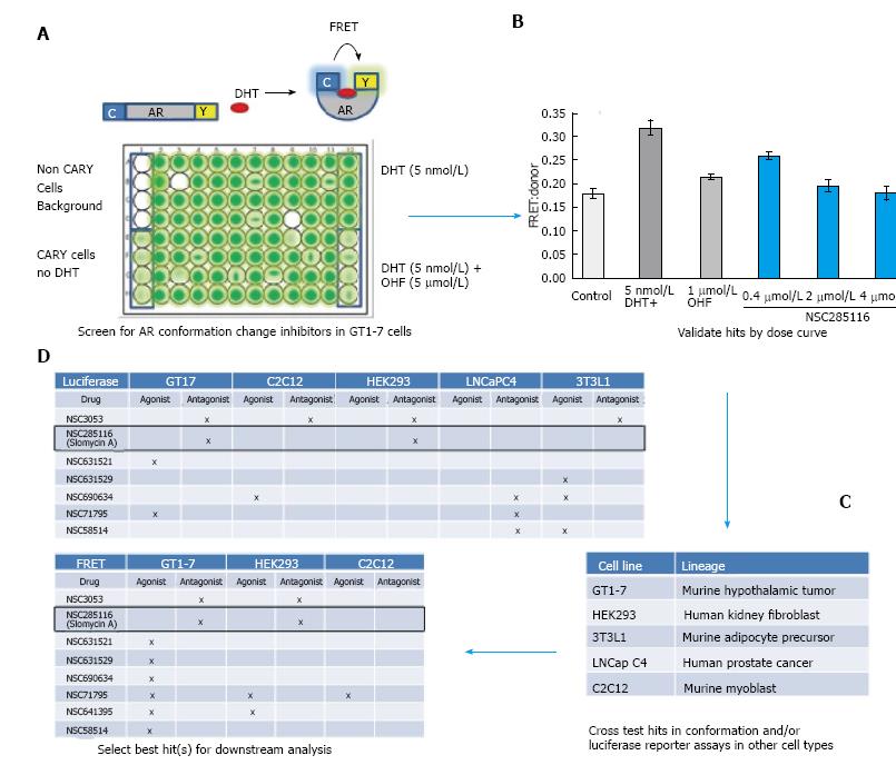
Figure 1 Screening for neuron-selective androgen receptor inhibitors.
A: AR conformation change can be monitored by the CFP-AR-YFP FRET reporter (top). Screening for compounds that affect AR conformation change in GT1-7 cells was accomplished in 96 well plates; B: Hits were assessed for a dose response in the same assay. Shown is the dose response to NSC285116 (siomycin A); C, D: Hits were then assessed for agonist or antagonist activity in the indicated cell lines in a FRET or an androgen-responsive luciferase reporter assay. Siomycin A demonstrated antagonist activity in GT1-7 neuron cells but not C2C12 muscle cells and was therefore selected for further analysis. AR: Androgen receptor.
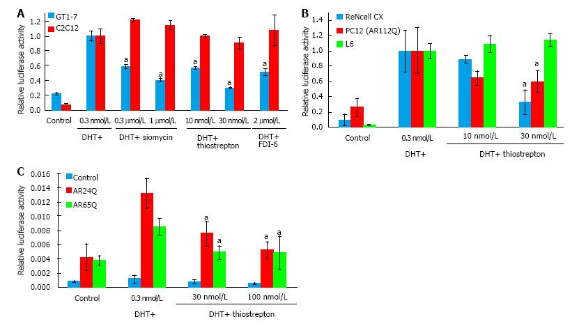
Figure 2 Thiazole antibiotics selectively inhibit androgen receptor in neuronal cells and have activity against polyQ-expanded androgen receptor.
A: GT1-7 and C2C12 cells were transfected with androgen-responsive and control luciferase reporters and treated with the indicated drugs overnight. Both thiazole antibiotics siomycin and thiostrepton, as well as the selective FOXM1 inhibitor FDI-6, inhibited AR activity in GT1-7 but not C2C12 cells, with thiostrepton being the most potent of the drugs; B: Rat L6 skeletal muscle cells and ReNcell CX and PC12 (ARQ112) neuronal cells were transfected with androgen-responsive and control luciferase reporters and treated with thiostrepton overnight. Thiostrepton inhibited AR luciferase activity in neuronal, but not muscle cells; C: 293 cells were transfected with androgen-responsive and control luciferase reporters as well as plasmids containing AR with 24 (normal state) or 65 (disease state) glutamines and treated with drugs mentioned above overnight. Both normal and disease state AR activity was reduced by thiostrepton treatment. Error bars represent the mean ± SEM, aP < 0.05. AR: Androgen receptor.
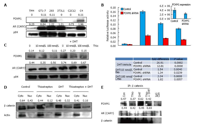
Figure 3 FOXM1 expression correlates with androgen receptor activity.
A: Expression of FOXM1 and AR was assessed by Western blot in indicated cell lines; B: GT1-7 cells were transfected with a lentiviral plasmid shRNA targeting FOXM1 as well as luciferase plasmids, treated with drugs listed above, then assessed by luciferase assay. Decreased expression of FOXM1 (RT-qPCR in inset) reduced AR activity and diminished sensitivity to thiostrepton. Fold changes and P values are shown; C: Western blot analysis demonstrates that thiostrepton reduces FOXM1 levels in GT1-7 cells while DHT increases FOXM1 levels; D: GT1-7 cells were treated with the indicated drugs overnight, and nuclear and cytoplasmic fractions were probed by Western blot for the indicated proteins. β-catenin levels in nuclear fraction of thiostrepton treated cells are lower compared to untreated ones. Quantification of β-catenin levels relative to cytoplasmic actin expression is shown; E: β-catenin was immunoprecipitated from GT1-7 cell lysates and Western blot was performed to detect AR and FOXM1. Quantification of AR and FOXM1 levels relative to β-catenin levels in IP is shown. AR: Androgen receptor.
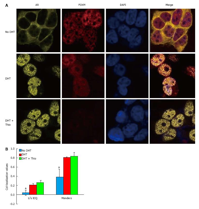
Figure 4 DHT promotes nuclear co-localization of androgen receptor and FOXM1.
A: GT1-7 cells expressing a YFP-tagged AR were treated as indicated for 24 h prior to staining with anti-FOXM1 (red) and DAPI (blue); B: Quantification of co-localization using Image J demonstrates significant co-localization of FOXM1 and AR in the presence of DHT and DHT + thiostrepton by both Manders split coefficient and Li’s ICQ value. Error bars represent the mean ± SEM, aP < 0.05. AR: Androgen receptor; ICQ: Intensity Correlation Quotient.
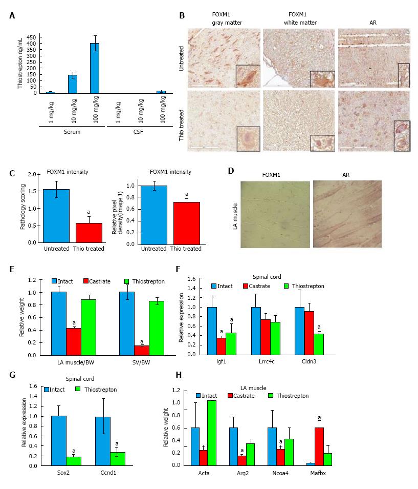
Figure 5 Thiostrepton treatment inhibits androgen receptor activity in rat spinal cord but not muscle.
A: Rats (n = 3) were administered 1, 10, or 100 mg/kg intraperitoneal injections thrice per week for four weeks. One hour following the final dose, blood and spinal fluid were collected and analyzed by mass spectrometry. Thiostrepton displayed the expected dose response in the serum, but much less was found in the cerebral spinal fluid; B-F: Rats (n = 7) were treated with 100 mg/kg per day thiostrepton or vehicle for four weeks using intraperitoneally implanted osmotic pumps. Intact and castrate cohorts were included as controls; B, C: Immunohistochemistry revealed a reduction in the expression of FOXM1 in spinal cords compared to untreated (intact or castrate) animals. AR staining was also strong in the neurons; C: FOXM1 staining intensity was quantified by a veterinary pathologist (left) or by ImageJ (right) and found to be significantly reduced by thiostrepton treatment; D: IHC revealed weak to no FOXM1 staining in LA muscle tissue while AR staining was prevalent; E: Tissues were harvested and weighed. Castration significantly reduced the weight of the LA muscle (P < 0.01) and seminal vesicles (SV) (P < 0.01) compared to intact animals while thiostrepton treatment did not. (F-H) RT-qPCR was used to quantify the levels of androgen-regulated transcripts in spinal cord (F) or LA muscle tissue (H) or FOXM1-regulated transcripts in spinal cord (G). Thiostrepton was able to decrease the levels of androgen-regulated transcripts in spinal cords to a similar or greater extent than castration, suggesting inhibition of AR activity, while in LA muscle, castration had a much stronger effect on AR activity than thiostrepton treatment. Two FOXM1 target gene transcripts were decreased with thiostrepton treatment, demonstrating an on target effect of the drug. Error bars represent the mean ± SEM, aP < 0.05 (P < 0.08 in G). AR: Androgen receptor.
- Citation: Otto-Duessel M, Tew BY, Vonderfecht S, Moore R, Jones JO. Identification of neuron selective androgen receptor inhibitors. World J Biol Chem 2017; 8(2): 138-150
- URL: https://www.wjgnet.com/1949-8454/full/v8/i2/138.htm
- DOI: https://dx.doi.org/10.4331/wjbc.v8.i2.138


