Copyright
©The Author(s) 2015.
World J Biol Chem. Aug 26, 2015; 6(3): 240-248
Published online Aug 26, 2015. doi: 10.4331/wjbc.v6.i3.240
Published online Aug 26, 2015. doi: 10.4331/wjbc.v6.i3.240
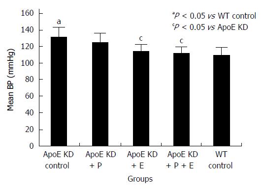
Figure 1 Effect of paricalcitol and enalapril alone and in combination for 16-wk on mean blood pressure (mmHg) in ApoE-deficient atherosclerotic mice.
There was a significant (P < 0.05) increase in mean BP in atherosclerotic mice compared to wild type control (n = 12). Enalapril (n = 12) alone and in combination with paricalcitol (n = 12) significantly (P < 0.05) reduced mean blood pressure (BP) in atherosclerotic mice (n = 12). Paricalcitol (n = 12) slightly but not significantly decreased mean BP in atherosclerotic mice.
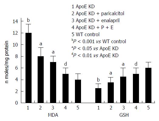
Figure 2 Effect of paricalcitol and enalapril alone and in combination for 16-wk on mono and combination for 16-wk on renal malondialdehyde and glutathione levels in ApoE-deficient atherosclerotic mice.
There was a significant (P < 0.001) increase in the renal MDA levels in atherosclerotic mice compared to wild type control (group 5). Paricalcitol (n = 6) and Enalapril (n = 6) alone significantly (P < 0.05 and P < 0.05) reduced aortic malondialdehyde (MDA) levels. However combination of the two (n = 6) decreased greater and significant (P < 0.01) aortic MDA levels than either drug alone in atherosclerotic mice (n = 6). There was a significant (P < 0.001) decrease in aortic glutathione (GSH) levels in atherosclerotic mice compared to wild type control (group 5). Paricalcitol (n = 6) and enalapril (n = 6) alone significantly (P < 0.05) increased aortic GSH levels. However combination of the two (n = 6) increased greater and significant (P < 0.01) aortic GSH levels than either drug alone in atherosclerotic mice (n = 6).
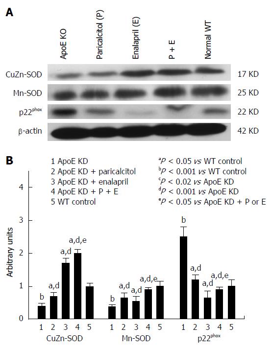
Figure 3 Western blot analysis and densitometry of protein band analysis to paricalcitol and enalapril alone and in combination for 16-wk on renal NADPH oxidase subunit p22phox, manganese-superoxide dismutase and copper/zinc-superoxide dismutase protein expression in ApoE-deficient atherosclerotic mice.
A: Western blot analysis of the effect of paricalcitol and enalapril alone and in combination for 16-wk on renal NADPH oxidase subunit p22phox, manganese-superoxide dismutase (Mn-SOD) and copper/zinc-superoxide dismutase (CuZn-SOD) protein expression in ApoE-deficient atherosclerotic mice; B: Densitometry of protein band analysis show that renal NADPH oxidase subunit p22phox and Mn-SOD expression significantly increased (P < 0.001) whereas CuZn-SOD protein expression significantly decreased (P < 0.001) in atherosclerotic mice (n = 3) compared to controls (n = 3). Paricalcitol (n = 3), enalapril (n = 3) and combination of the two (n = 3) significantly (P < 0.02 and P < 0.001) ameliorated the oxidative stress by inhibiting the induction of NADPH oxidase and Mn-SOD expression (38%, 76% and 68%, respectively) and up-regulating the CuZn-SOD protein expression (52%, 342% and 398%, respectively) in atherosclerotic mice. The data represent mean ± SE of three independent Western blot experiments.
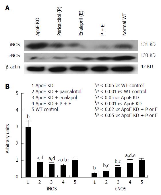
Figure 4 Western blot analysis and densitometry of protein band analysis to paricalcitol and enalapril alone and in combination for 16-wk on renal iNOS and eNOS protein expression in ApoE-deficient atherosclerotic mice.
A: Western blot analysis of the effect of paricalcitol and enalapril alone and in combination for 16-wk on renal iNOS and eNOS protein expression in ApoE-deficient atherosclerotic mice; B: Densitometry of protein band analysis show that renal iNOS expression was significantly enhanced (P < 0.001) whereas eNOS protein expression was significantly decreased (P < 0.001) in ApoE-deficient atherosclerotic mice (n = 3) compared to controls (n = 3). Pricalcitol (n = 3), enalapril (n = 3) and the combination of the two (n = 3) significantly (P < 0.05 and P < 0.001) ameliorated the alterations in iNOS and eNOS expression in atherosclerotic mice. The data represent mean ± SE of three independent Western blot experiments.
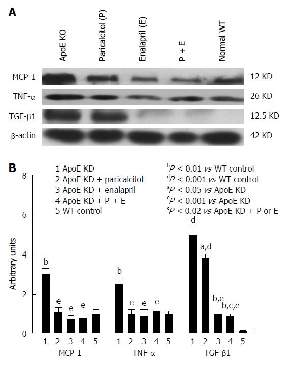
Figure 5 Western blot analysis and densitometry of protein band analysis to paricalcitol and enalapril alone and in combination for 16-wk on renal inflammatory proteins MCP-1, TNF-α and TGF-β1 expression in ApoE-deficient atherosclerotic mice.
A: Western blot analysis of the effect of paricalcitol and enalapril alone and in combination for 16-wk on renal inflammatory proteins MCP-1, TNF-α and TGF-β1 expression in ApoE-deficient atherosclerotic mice; B: Densitometry of protein band analysis show that renal MCP-1, TNF-α and TGF-β1 expression was significantly enhanced (P < 0.01, P < 0.01 and P < 0.001) in ApoE-deficient atherosclerotic mice (n = 3) compared to controls (n = 3). Pricalcitol (n = 3) and enalapril (n = 3) alone and in combination of the two (n = 3) significantly (P < 0.05 and P < 0.001) exerted anti-inflammatory response by down-regulating the MCP-1, TNF-α and COX-2 expression in atherosclerotic mice. The data represent mean ± SE of three independent Western blot experiments.
- Citation: Husain K, Suarez E, Isidro A, Hernandez W, Ferder L. Effect of paricalcitol and enalapril on renal inflammation/oxidative stress in atherosclerosis. World J Biol Chem 2015; 6(3): 240-248
- URL: https://www.wjgnet.com/1949-8454/full/v6/i3/240.htm
- DOI: https://dx.doi.org/10.4331/wjbc.v6.i3.240


