Copyright
©2014 Baishideng Publishing Group Inc.
World J Biol Chem. Nov 26, 2014; 5(4): 457-464
Published online Nov 26, 2014. doi: 10.4331/wjbc.v5.i4.457
Published online Nov 26, 2014. doi: 10.4331/wjbc.v5.i4.457
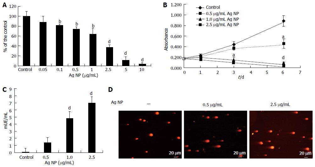
Figure 1 Silver nanoparticles are cytotoxic and genotoxic for human dermal microvascular endothelial cells.
(A) Human dermal microvascular endothelial cells (HMEC) were exposed to various concentrations of silver nanoparticles (Ag NP) for 72 h or (B) were treated with different concentrations of Ag NP for different times. MTT assay was then performed. In (A) data are expressed as the percentage of control and in (B) as absorbance. Data represent the mean ± SD deviation of at least three separate experiments in triplicate. P value was calculated vs untreated cells: aP < 0.05, bP < 0.01, dP < 0.001. C: HMEC were treated with 0.5, 1.0 and 2.5 μg/mL Ag NP for 24 h and the LDH leakage assay was performed. Data are expressed as enzyme unit/mL culture medium and represent the mean ± SD of five separate experiments. P value was calculated vs untreated cells: dP < 0.001; D: Comet assay was performed on HMEC treated with Ag NP for 24 h. After staining with ethidium bromide, the slides were analyzed with a fluorescence microscope.
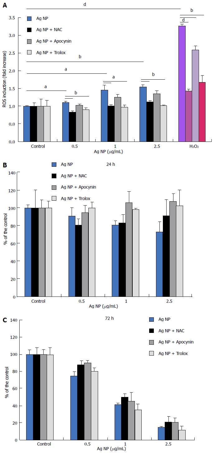
Figure 2 Silver nanoparticles induce the formation of reactive oxygen species, but antioxidants do not prevent silver nanoparticles’ cytotoxicity.
A: HMEC cells were pretreated with antioxidants and then exposed to various concentrations of Ag or H2O2 (100 μmol/L) for 30 min. ROS generation was then measured. Data are shown as the fold increase in ROS levels of treated cells compared to the corresponding control. Each bar is the mean of three separate experiments ± SD. P value was calculated vs untreated cells: aP < 0.05, bP < 0.01, dP < 0.001; B: HMEC were pre-incubated with Trolox (40 μmol/L), NAC (5 mmol/L) or apocynin (10 μmol/L) for 2 h before adding different concentrations of Ag NP. MTT assay was performed after 24 h (B) and 72 h (C). Data are expressed as the percentage of control and represent the mean ± SD of four separate experiments. HMEC: Human dermal microvascular endothelial cells.
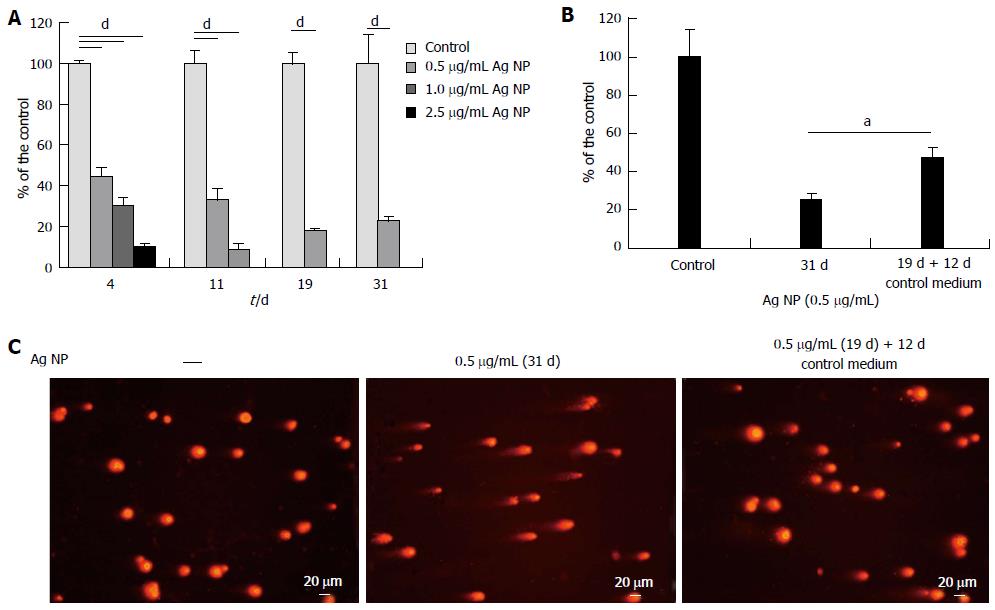
Figure 3 Low concentrations of silver nanoparticles reversibly retard human dermal microvascular endothelial cells growth.
A: Human dermal microvascular endothelial cells (HMEC) were exposed to various concentrations of silver nanoparticles (Ag NP). All the samples were trypsinized and counted on the same day in which the controls reached confluence. After counting, the cells were re-seeded at the same density; B: On day 19, part of the samples were seeded in the absence of Ag NP and counted at day 31. In (A) and (B) data are expressed as the percentage of control. Data represent the means ± SD of four separate experiments. In (A) P value was calculated vs untreated cells, dP < 0.001; and in (B) P value was calculated vs cells cultured for 31 d continuously with Ag NP, aP < 0.05; C: Comet assay was performed as described on HMEC treated with Ag NP (0.5 μg/mL) for 31 d or after removal of the nanoparticles from the culture media on day 19.
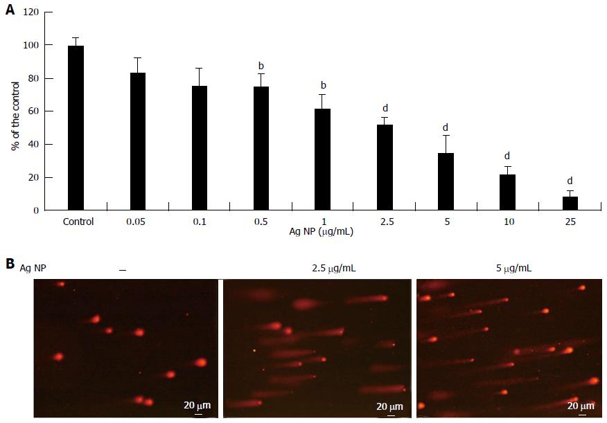
Figure 4 Silver nanoparticles are cytotoxic and genotoxic for endothelial colony-forming cells.
A: Endothelial colony-forming cells (ECFC) were treated with various concentrations of silver nanoparticles (Ag NP) for 72 h. MTT assay was then performed and data are expressed as the percentage of control. Data represent the mean ± SD of at least three separate experiments. P value was calculated vs untreated cells: bP < 0.01, dP < 0.001. B: Comet assay was performed on ECFC treated with Ag NP (2.5 and 5 μg/mL) for 24 h as described.
- Citation: Castiglioni S, Caspani C, Cazzaniga A, Maier JA. Short- and long-term effects of silver nanoparticles on human microvascular endothelial cells. World J Biol Chem 2014; 5(4): 457-464
- URL: https://www.wjgnet.com/1949-8454/full/v5/i4/457.htm
- DOI: https://dx.doi.org/10.4331/wjbc.v5.i4.457


