Copyright
©2011 Baishideng Publishing Group Co.
World J Biol Chem. Feb 26, 2011; 2(2): 35-38
Published online Feb 26, 2011. doi: 10.4331/wjbc.v2.i2.35
Published online Feb 26, 2011. doi: 10.4331/wjbc.v2.i2.35
Figure 1 Lee G Pedersen, PhD, Professor, Department of Chemistry, University of North Carolina at Chapel Hill, CB#3290, Chapel Hill, NC 27599, United States.
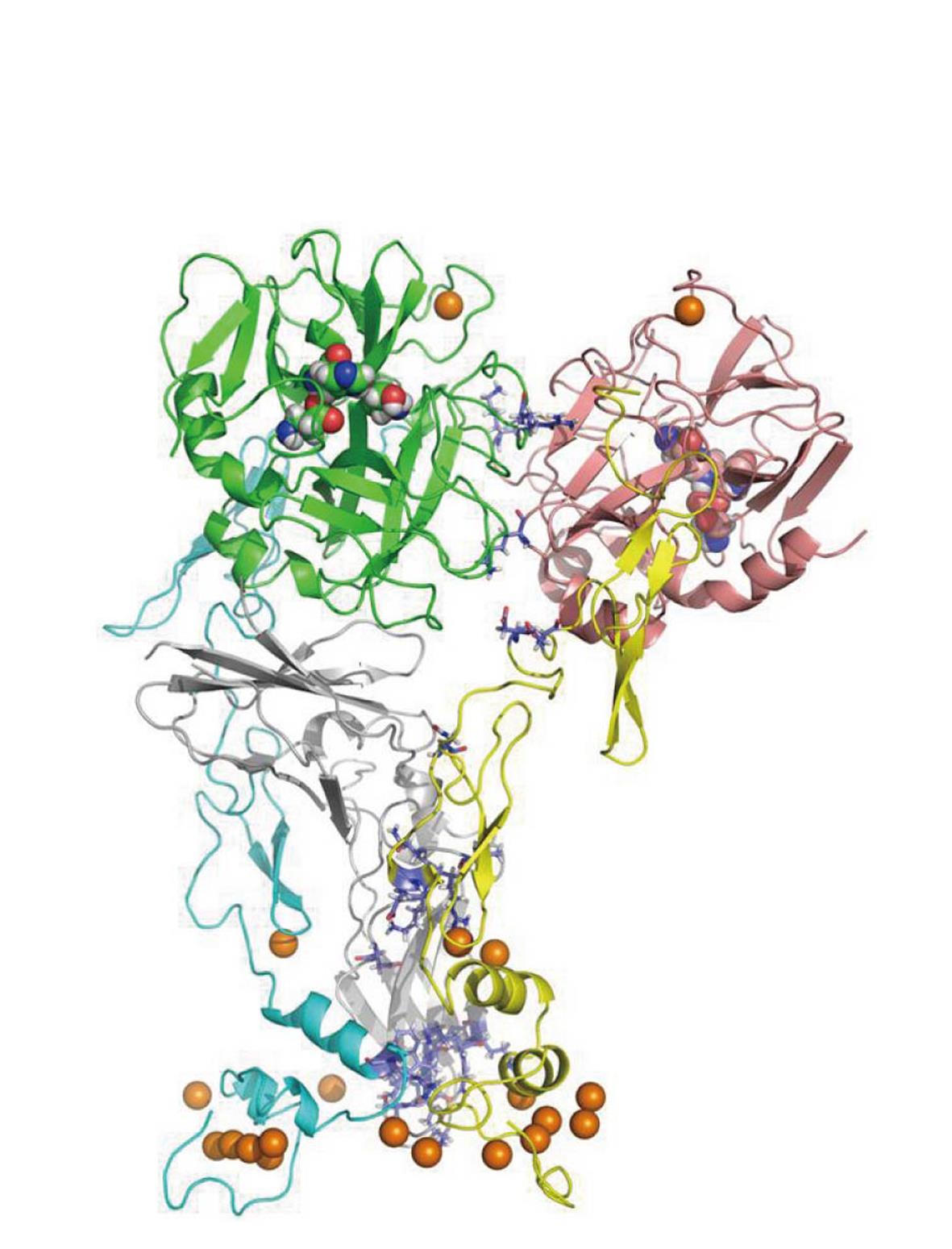
Figure 2 A solvent-equilibrated model of human factor VIIa/tissue factor/factor Xa.
Left: Factor VIIa (FVIIa)-the SP domain is green, while the EGF2-EGF1-GLA domains are blue. Right: FXa-the SP domain is maroon, while the EGF2-EGF1-GLA domains are yellow. Center: Tissue factor-the two domains are shown in dark gray. The gold colored spheres are Ca2+ ions. The three residues that define the active sites of the serine protease domains are shown by space filling atoms. There is no experimental structure to date for this complex.
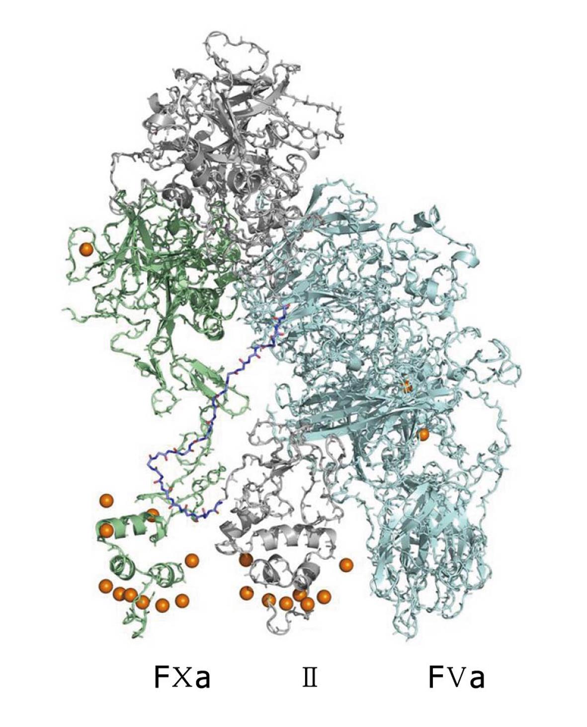
Figure 3 A solvent-equilibrated model of human factor Va/factor Xa/II.
factor Xa (FXa) is on the left in green, FVa is on the right in blue and II (prothrombin) is in the center in gray. There is no experimental structure to date for this complex
- Citation: Pedersen LG. Lee Pedersen’s work in theoretical and computational chemistry and biochemistry. World J Biol Chem 2011; 2(2): 35-38
- URL: https://www.wjgnet.com/1949-8454/full/v2/i2/35.htm
- DOI: https://dx.doi.org/10.4331/wjbc.v2.i2.35


