Copyright
©2011 Baishideng Publishing Group Co.
World J Biol Chem. Oct 26, 2011; 2(10): 232-238
Published online Oct 26, 2011. doi: 10.4331/wjbc.v2.i10.232
Published online Oct 26, 2011. doi: 10.4331/wjbc.v2.i10.232
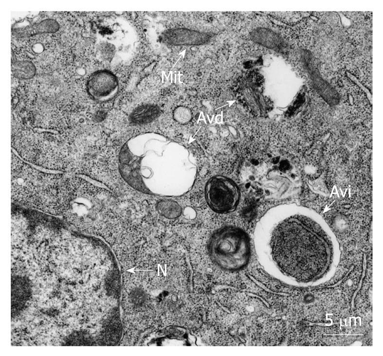
Figure 1 Morphology of autophagic vacuoles.
Typical autophagic vacuoles from 3T3 mouse fibroblasts incubated in a nutrient-poor medium containing cytoplasmic material at early (Avi) and late (Avd) degradation stages. Mit: Mitochondria; N: Nucleus.
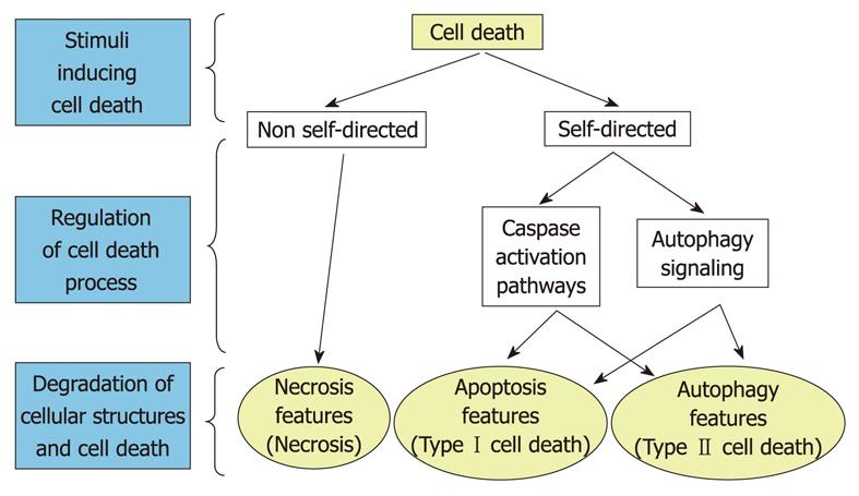
Figure 2 Main forms of cell death.
Autophagy is located in the proper context in relation to classical necrosis and apoptosis. Crossing arrows indicate the existence of common links for apoptosis and autophagy. The entire process of cell death has been divided into three phases: stimulation, regulation and degradation. Note the absence of a regulation phase in necrosis.
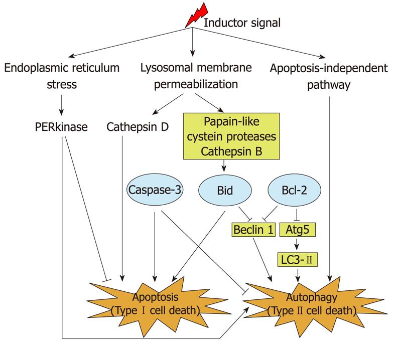
Figure 3 Model of regulation of autophagy and apoptosis in breast cancer cells.
Different molecules described to act as a link between apoptosis and autophagy are shown (see text for details). Ellipses indicate classical regulators of apoptosis. Moreover, organelles, such as mitochondria, endoplasmic reticulum and lysosomes, appear to be involved in this regulation. Arrow-headed lines and bar-headed lines indicate activation and inhibition, respectively, of their corresponding targets.
- Citation: Esteve JM, Knecht E. Mechanisms of autophagy and apoptosis: Recent developments in breast cancer cells. World J Biol Chem 2011; 2(10): 232-238
- URL: https://www.wjgnet.com/1949-8454/full/v2/i10/232.htm
- DOI: https://dx.doi.org/10.4331/wjbc.v2.i10.232


