Copyright
©2010 Baishideng Publishing Group Co.
World J Biol Chem. May 26, 2010; 1(5): 188-195
Published online May 26, 2010. doi: 10.4331/wjbc.v1.i5.188
Published online May 26, 2010. doi: 10.4331/wjbc.v1.i5.188
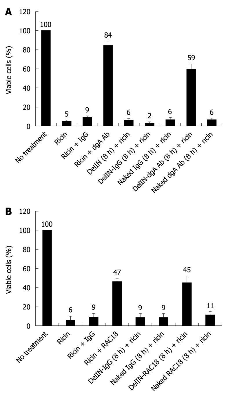
Figure 1 Protective effects of Ab-DeliverIN (DelIN)-mediated intracellular delivery of polyclonal anti-deglycosylated ricin A-chain antibody (anti-dgA Ab) (A) and monoclonal RAC18 anti-ricin A-chain monoclonal antibody (RAC18 mAb) (B) against ricin-induced cytotoxicity.
RAW264.7 cells were incubated with naked antibodies (dgA Ab, RAC18 mAb or control IgG) (no liposome encapsulation) or DelIN-encapsulated antibodies (DelIN-dgA Ab, DelI-RAC18 mAb or DelIN-control IgG) for 8 h. After 3 washes with PBS to remove extracellular antibodies, ricin (100 ng/mL) was added and incubated for additional 24 h. Cell viability was quantitated using the MTT assay. As controls for the neutralizing effects of anti-ricin antibodies, ricin was co-incubated with naked dgA Ab, RAC18 mAb or control IgG.
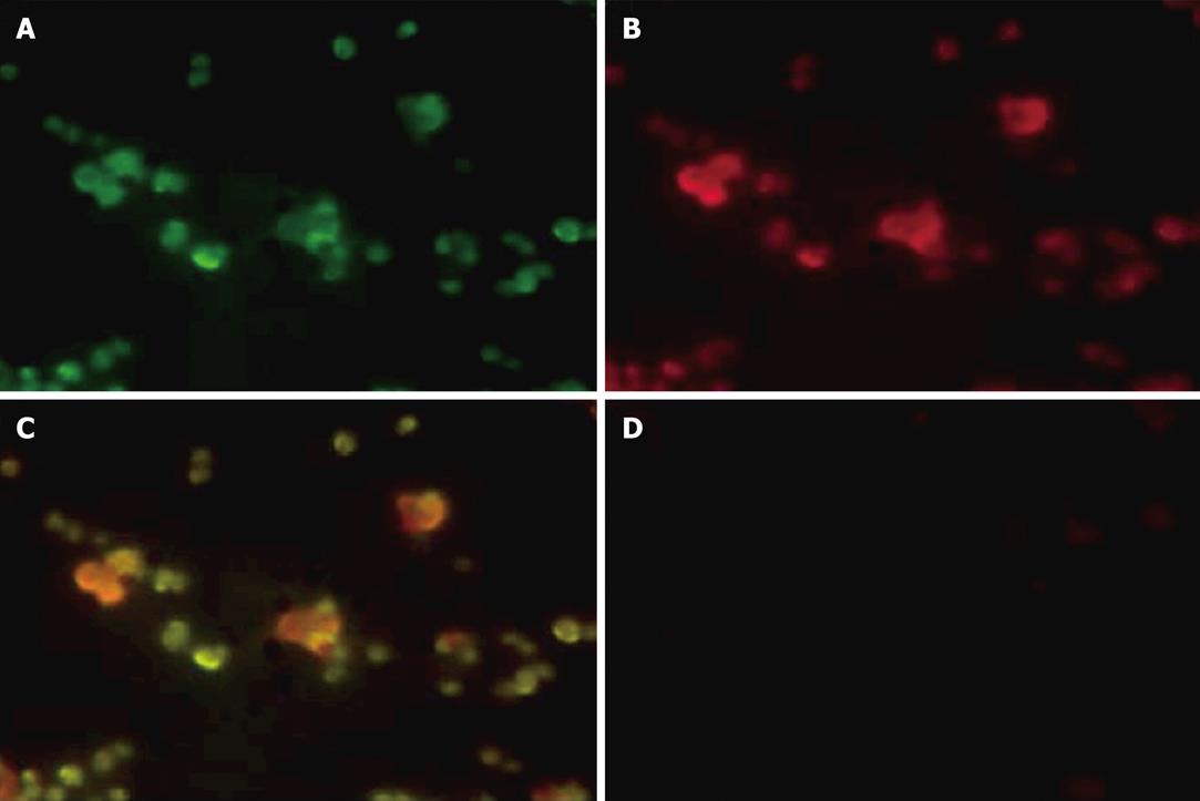
Figure 2 Intracellular localization of fluorescent-labeled ricin and fluorescent-labeled DelIN-encapsulated RAC18 mAb by immunofluorescent staining.
Immunofluorescent staining of RAW264.7 cells treated with Alexa 488-labeled ricin and DelIN-encapsulated Alexa 594-labeled RAC18 mAb. A: Alexa 488-labeled ricin (green); B: DelIN-Alexa 594-labeled RAC18 mAb (red); C: Superposition of A and B demonstrated co-localization of ricin and RAC18 mAb (yellow); D: Immunofluorescent staining of RAW264.7 cells treated with naked Alexa 594-labeled RAC18 mAb. No staining was observed, demonstrating the inability of naked RAC18 mAb to enter cells.
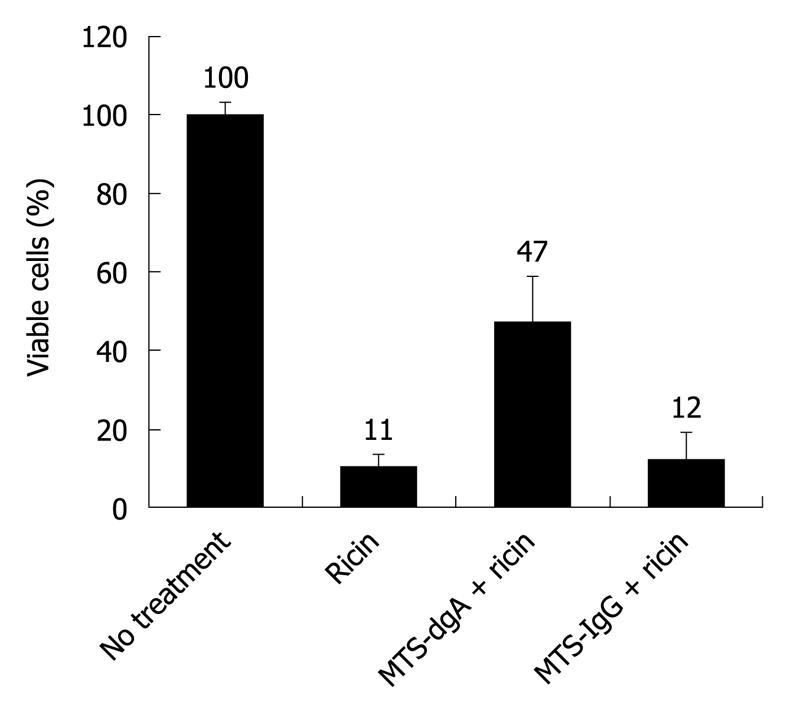
Figure 3 Neutralizing effect of MTS-conjugated dgA Ab against ricin-induced cytotoxicity.
To confirm the neutralizing activity of dgA Ab conjugated to the MTS cell-penetrating peptide, RAW264.7 cells were co-incubated with ricin and MTS conjugated to dgA Ab (MTS-dgA Ab) or MTS-conjugated to control IgG (MTS-IgG) for 24 h. Cell viability was quantitated using the MTT assay.
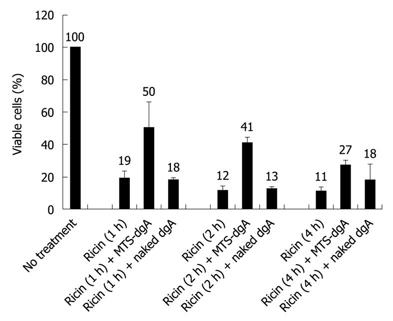
Figure 4 Protective effects of MTS-conjugated dgA Ab for post-exposure treatment of ricin-induced cytotoxicity.
RAW264.7 cells were exposed to ricin (100 ng/mL) for 1 h, 2 h or 4 h to allow time for the toxin to internalize into cells. After a short exposure, cells were washed 3 times with PBS to remove extracellular ricin. Then MTS-conjugated dgA antibody or naked (unconjugated) dgA Ab (1 μg/well) was added and incubated for another 24 h and cell viability was quantitated by the MTT assay. Naked dgA Ab, which could not enter cells, served as a negative control for the neutralizing effects of MTS-conjugated dgA Ab.
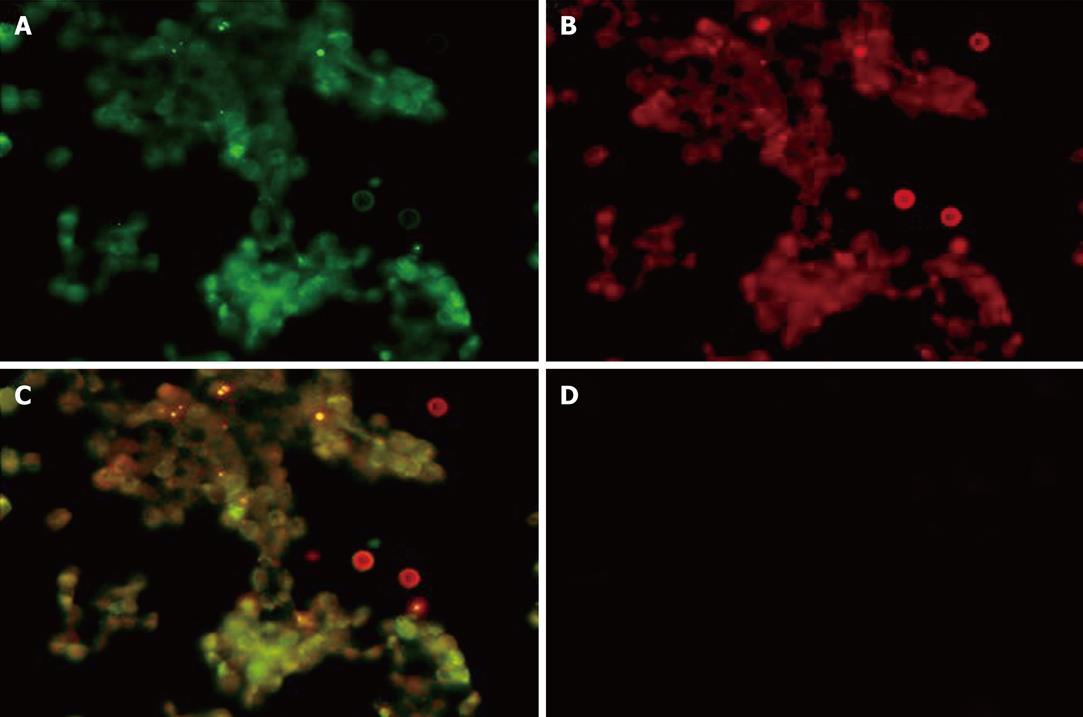
Figure 5 Intracellular localization of fluorescent-labeled ricin and MTS-conjugated dgA Ab by immunofluorescent staining.
A: Immunofluorescent staining of RAW264.7 cells treated with Alexa 488-labeled ricin (green); B: MTS-conjugated dgA Ab detected by TRITC-labeled anti-mouse IgG (red); C: Superposition of A and B demonstrated co-localization of ricin and MTS-dgA Ab (yellow); D: Immunofluorescent staining of RAW264.7 cells treated with unconjugated dgA Ab and detected with TRITC-labeled anti-mouse IgG. No staining was observed, demonstrating the inability of unconjugated dgA Ab to enter cells.
- Citation: Wu F, Fan S, Martiniuk F, Pincus S, Müller S, Kohler H, Tchou-Wong KM. Protective effects of anti-ricin A-chain antibodies delivered intracellularly against ricin-induced cytotoxicity. World J Biol Chem 2010; 1(5): 188-195
- URL: https://www.wjgnet.com/1949-8454/full/v1/i5/188.htm
- DOI: https://dx.doi.org/10.4331/wjbc.v1.i5.188


