Published online Dec 27, 2017. doi: 10.4240/wjgs.v9.i12.264
Peer-review started: August 7, 2017
First decision: September 6, 2017
Revised: November 6, 2017
Accepted: November 19, 2017
Article in press: November 19, 2017
Published online: December 27, 2017
Processing time: 142 Days and 21 Hours
To study the utility of single-incision totally extraperitoneal inguinal hernia repair with intraperitoneal inspection.
A 2 cm transverse skin incision was made in the umbilicus, extending to the intraperitoneal cavity. Carbon dioxide was insufflated followed by insertion of laparoscope to observe the intraperitoneal cavity. The type of hernia was diagnosed and whether there was the presence of intestinal incarceration was confirmed. When an intestinal incarceration in the hernia sac was found, the forceps were inserted through the incision site and the intestine was returned to the intraperitoneal cavity without increasing the number of trocars. Once the peritoneum was closed, totally extraperitoneal inguinal hernia repair was performed, and finally, intraperitoneal observation was performed to reconfirm the repair.
Of the 75 hernias treated, 58 were on one side, 17 were on both sides, and 10 were recurrences. The respective median operation times for these 3 groups of patients were 100 min (range, 66 to 168), 136 min (range, 114 to 165), and 125 min (range, 108 to 156), with median bleeding amounts of 5 g (range, 1 to 26), 3 g (range, 1 to 52), and 5 g (range, 1 to 26), respectively. Intraperitoneal observation showed hernia on the opposite side in 2 cases, intestinal incarceration in 3 cases, omental adhesion into the hernia sac in 2 cases, severe postoperative intraperitoneal adhesions in 2 cases, and bladder protrusion in 1 case. There was only 1 case of recurrence.
Single-incision totally extraperitoneal inguinal hernia repair with intraperitoneal inspection makes hernia repairs safer and reducing postoperative complications. The technique also has excellent cosmetic outcomes.
Core tip: Single-incision totally extraperitoneal inguinal hernia repair with intraperitoneal inspection (iSTEP) makes hernia repairs safer and more effectively. Totally extraperitoneal inguinal hernia repair had the disadvantages for difficulty with confirming the type of hernia as well as difficulty with large indirect inguinal hernia, intestinal incompetence and postoperative prostatectomy. However, iSTEP can be used to diagnose the type of hernia easily. It enables observation of the opposite side and reconfirmation of treatment after mesh repair making the technique safer and reducing postoperative complications. The technique also has excellent cosmetic outcomes.
- Citation: Yamamoto M, Urushihara T, Itamoto T. Utility of single-incision totally extraperitoneal inguinal hernia repair with intraperitoneal inspection. World J Gastrointest Surg 2017; 9(12): 264-269
- URL: https://www.wjgnet.com/1948-9366/full/v9/i12/264.htm
- DOI: https://dx.doi.org/10.4240/wjgs.v9.i12.264
Prudent observation of inguinal hernia is recommended for asymptomatic patients according to the guideline for inguinal hernia treatment released by the European Hernia Society[1]. However, surgery is the standard treatment. The surgical techniques used for inguinal hernia consist of the traditional anterior technique and laparoscopic surgery. Two approaches are utilized in laparoscopic surgery: Transabdominal preperitoneal approach (TAPP) and totally extraperitoneal hernia repair (TEP). In cases of intestinal incarceration, intraperitoneal observation may be necessary to confirm the presence of intestinal damage after reduction, but many inguinal hernias can be repaired without entering the abdominal cavity. TEP shortens the operation time, enhances patient satisfaction, and reduces postoperative pain[2]. However, using conventional TEP, only the hernia on one side can be identified, and there is a possibility of missing occulting inguinal hernia on the opposite side. By combining TEP with intraperitoneal observation, it is possible to diagnose the type of hernia, confirm repair after covering the hernia with mesh, and perform both procedures safely and reliably. We report herein our experiences with single-incision totally extraperitoneal inguinal hernia repair with intraperitoneal inspection (iSTEP), which was performed on 75 patients.
From April 2009 when iSTEP was first introduced until May 2016, the 75 patients who underwent the procedure at the Prefectural Hiroshima Hospital were enrolled. All surgeries were performed by the same experienced surgeon. Data on patient demographics, clinical data, intraoperative findings, and postoperative course were prospectively collected. All patients underwent surgery after providing informed consent. The procedure was approved by the Ethics Committee at the Prefectural Hiroshima Hospital and the study was performed in accordance with the Declaration of Helsinki. We excluded patients who met the following criteria: History of prostate surgery, giant inguinal hernia, young patients with small indirect inguinal hernia, strangulated hernia, and patients who could not tolerate general anesthesia, which was employed for laparoscopic hernia repair at our hospital.
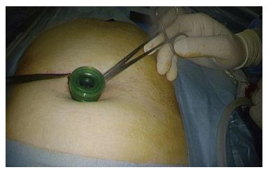
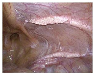
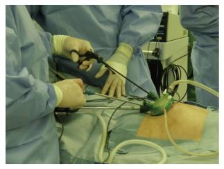
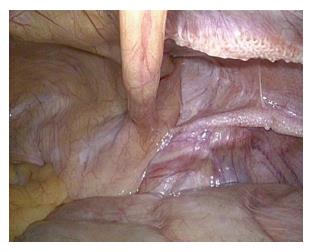
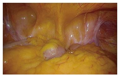
During the surgery, the patient was placed in a supine position under general anesthesia. A 2 cm transverse skin incision was made in the umbilicus, followed by an incision in the peritoneum from the fascia defect to the abdominal cavity. A trocar attached to an access port was inserted and carbon dioxide was insufflated to 8 mmHg (Figure 1). The type of hernia was diagnosed and the presence of intestinal incarceration was confirmed in the intraperitoneal cavity (Figure 2). The trocar was removed and the peritoneum was closed after inserting a catheter to degas the cavity. The peritoneum was ligated by 3-0 Vicryl, once the peritoneum was closed (the ligation was unfolded at the time performing intraperitoneal observation). TEP was then started. The subcutaneous tissue was dissected to the rectus abdominis anterior sheath and a transecting incision was made at the anterior sheath. The rectus abdominis was split and the posterior sheath was exposed. Blunt dissection using an electrical scalpel or a finger was performed between the muscle and the posterior sheath to create a preperitoneal space. A multi-channel access port (GelPOINT MINI; Applied Medical, Rancho Santa Margarita, CA, United States) was installed and carbon dioxide was insufflated to 8mmHg again (Figure 3). The preperitoneal space was dissected using a bipolar forceps by grasping the forceps and pulling toward the Retzius cavity and the peritoneal edge was checked. The cord structures were freed from the hernia sac and parietalisation was performed gently without perforation of the peritoneum. The hernia sac was extracorporeally ligated with a Fisherman’s knot using 2-0 Prolene and dissected. The edge of the peritoneum was grasped and dissected toward the dorsal side and the lateral side to secure a space for mesh. The Gel Seal CAP was detached and an artificial patch (3D Max light Mesh L size; Bard, Murray Hill, NJ, United States) (10.8 cm × 16 cm) or TiLENE Mesh; pfm medical, Koln, Germany) (10 cm × 15 cm) was inserted through the incision. After the mesh was positioned to cover the Hesselbach triangle, femoral rings, and inguinal ring, it was fixed to the Cooper’s ligaments. The interior and lateral sides of the mesh were secured using a tracking device (Pro Tack; Medtronic, Fridley, MN, United States), applied carefully to avoid injury to the inferior epigastric vessels. Finally, the abdominal cavity was observed to confirm that the repair was complete (Figure 4). The peritoneum, anterior rectus sheath and skin were each closed.
iSTEP hernia repair was successfully completed in 75 patients. Patient demographics and characteristics are summarized in Table 1. There were 66 men and 9 women. The median age of the patients was 68 years (range, 17 to 82 years), median weight was 63 kg (range, 38 to 106 kg), and median body mass index was 23.0 kg/m2 (17.3 to 32.7 kg/m2). The number of patients with a physical status of ASA I, II, and III according to the American Society of Anesthesiologists classification was 25, 49, and 1, respectively. The subjects included smokers, individuals with hypertension, diabetes, respiratory disease, coronary artery disease, or taking anticoagulant/antiplatelet medicine. Fifty-eight hernias were on one side, 17 were on both sides, and 10 were recurrences. The median operation time for these 3 groups of patients was 100 minutes (range, 66 to 168), 136 min (range, 114 to 165), and 125 min (range, 108 to 156) and the median bleeding amount was 5 g (range, 1 to 26), 3 g (range, 1 to 52), 5 g (range, 1 to 26), respectively (Table 2). Intraperitoneal observation showed hernia on the opposite side in 2 cases, intestinal incarceration in 3 cases, omental adhesion to the hernia sac in 2 cases, severe postoperative intraperitoneal adhesions in 2 cases, and bladder protrusion in 1 case. Postoperative hemorrhage and wound infection were not observed, and there was only 1 case of recurrence.
| Variable | n (%) |
| Number of patients | 75 |
| Male | 66 (88) |
| Female | 9 (12) |
| Median age, yr (range) | 68 (17-82) |
| Median body weight, kg (range) | 63 (17.3-32.7) |
| Median BMI, kg/m2 (range) | 23.0 (17.3-32.7) |
| ASA score | |
| I | 25 (33.3) |
| II | 49 (65.3) |
| III | 1 (1.4) |
| Site of hernia | |
| Right | 33 (44) |
| Left | 25 (33.3) |
| Both | 17 (22.7) |
| Smoking | 37 (49.3) |
| Hypertension | 26 (34.7) |
| Diabetes mellitus | 7 (9.3) |
| Respiratory disease | 10 (13.3) |
| Coronary artery disease | 6 (8) |
| Anticoagulant/antiplatelet medicine | 7 (9.3) |
| Variable | Value | % |
| Operative time | ||
| Unilateral (min) | 100 (66-168) | |
| Bilateral (min) | 136 (114-165) | |
| Recurrence (min) | 125 (108-156) | |
| Bleeding volume (mL) | 4 (1-52) | |
| Conversion to multi-port or open | 0 | 0 |
| Intraoperative complication | 0 | 0 |
| Postoperative complication | ||
| Seroma | 0 | 0 |
| Wound infection | 0 | 0 |
| Chronic pain | 0 | 0 |
| Recurrence | 1 | 1.4 |
Compared to the conventional anterior approach, TEP results in less postoperative pain, fewer postoperative complications, lower recurrence rates, early discharge, and faster return to daily life[3]. TEP is classified as Level 1A treatment in the European hernia guidelines[1]. Moreover, it has a superior cosmetic outcome as it is performed through single-incision laparoscopic surgery[4]. Coupled with intraperitoneal observation, it is possible to diagnose the type of hernia, restore the intestinal tract incompetence and confirm the repair afterward. Hernia repairs can therefore be performed more safely and effectively.
A minimally invasive surgical technique for the repair of inguinal hernia, TEP was introduced for laparoscopic hernia repair in the early 1990s[5] and many studies involving the procedure have been reported since. Advantages of TEP include a wide range of exfoliation, ease of mesh placement, short operation time, and no need to perform peritoneal closure. Furthermore, by conducting TEP with single-incision laparoscopic surgery, it is possible to obtain excellent cosmetic outcomes as reported by Filipovic-Cugura et al[6] in 2009. However, the disadvantages of the procedure include difficulty with confirming the type of hernia as well as difficulty with large indirect inguinal hernia, intestinal incompetence and postoperative prostatectomy.
We complemented STEP with intraperitoneal observation to compensate for these drawbacks and obtained good results. Although the operation time is longer than that of the conventional procedure and the multiport laparoscopic surgery, the bleeding volume is equivalent, and the outcome is excellent with respect to postoperative complications[7-9]. Cost is also equal because special equipment is not required. Furthermore, compared to single-incision TAPP, STEP is easier in terms of exfoliation and thus the operation time can be shortened[2]. For patients with recurrent hernia, however, the surgical time was longer because of difficulties with the exfoliation procedure, but there was no conversion to TAPP at our hospital.
By using intraperitoneal observation in combination with STEP, it is possible to view the inguinal region without overlooking coexisting lesions, for example, the presence of hernia on the opposite side (Figure 5). Through intraperitoneal observation, it is possible to confirm the mesh coverage in cases where the hernia extends not only to direct and indirect inguinal lesions but also to femoral and obturator lesions. We could confirm the herniated gates extended to the femoral and obturator in 2 of our patients and could perform the necessary repairs reliably. A small hernia was also detected on the opposite side in some of the patients and this was treated simultaneously by performing hernia repair on both sides. In recurrent cases, iSTEP facilitates reliable repair due to reliable identification of the hernia gates.
In addition, if an intestinal incarceration in the hernia sac was found in the intraperitoneal cavity, forceps were inserted through the incision site and the intestine was returned to the abdominal cavity without increasing the number of trocars. Moreover, the presence of intestinal damage could be confirmed. The intestine protruded into the sac in 3 patients. In all 3 cases, we returned the intestine to the abdominal cavity, confirmed that there was no damage, and then performed TEP safely. If it was difficult to perform hernia repair using TEP, we could easily switch to TAPP. We used TAPP for patients that underwent prostate surgery and had severe adhesion in the preperitoneal space. However, we used TEP for 2 patients for whom TAPP would have been difficult due to severe adhesion in the abdominal cavity after abdominal surgery. Since the mesh is located in the preperitoneal space, even if there is adhesion in the abdominal cavity, the adhesion causes no issues during surgery and the risk of organ damage is low.
In cases of large direct inguinal hernia, it is difficult to identify the hernia gate using the anterior approach and it is difficult to dissect the medial and ventral sides using TAPP. Both sides can be dissected easily with TEP. When using the mesh recommended by the European Hernia Society, which is 15 cm × 10 cm in size[1], it is necessary to secure a sufficient dissection range, which is easy to do with TEP. In addition, peritoneal closure is difficult when performing TAPP through a single-incision procedure. Compared to TAPP, the advantages of TEP are that it does not require intraperitoneal manipulation and adhesion exfoliation can be omitted.
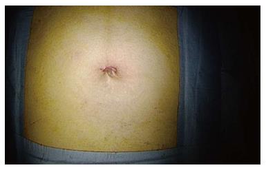
Postoperatively, there is a risk of bowel obstruction caused by intraperitoneal operation using TAPP and adhesion at the peritoneal closure region. The risk of intestinal obstruction after inguinal hernia repair using TAPP or TEP is 2.8 and 0.6 times the risk using the Lichtenstein method, respectively[10]. Additionally, by observing the interior of the abdominal cavity again after hernia repair, it can be confirmed that the fragile portion is covered by mesh and the risk of recurrence can be reduced. By conducting hernia repair with single-incision laparoscopic surgery, we could obtain excellent cosmetic outcomes (Figure 6). As demonstrated above, STEP with intraperitoneal inspection is a very useful technique because diagnosis and reinforcement can be performed reliably and the cosmetic outcome is excellent.
The present study has several limitations. First, this study was carried out at a single high-volume center and was retrospective in nature; hence, patient selection bias may have been inevitable. Patients who met the exclusion criteria were excluded. Second, the population number was small. Further studies on a larger scale are necessary.
Surgery is the standard treatment for inguinal hernia. The surgical techniques used for inguinal hernia consist of the traditional anterior technique and laparoscopic surgery. One type of laparoscopic surgery has totally extraperitoneal hernia repair (TEP). The outcome of TEP is superior to the conventional anterior approach; less postoperative pain, fewer postoperative complications, lower recurrence rates, early discharge, and faster return to daily life and superior cosmetic outcome. The authors cannot observe intraperitoneal cavity on TEP, so the opposite side hernia might be overlooked if the hernia is present. And it is difficult to perform the procedure for patients with intestinal incarceration in the hernia sac.
It is difficult to repair hernia by TEP for patients with large indirect inguinal hernia, intestinal incarceration and postoperative prostatectomy. The authors must compensate for these drawbacks of TEP.
By using intraperitoneal inspection (iSTEP), it is possible to view the inguinal region without overlooking coexisting lesions if the hernia present on the opposite side. And when an intestinal incarceration in the hernia sac was found, we can return the intestine and confirm the presence of intestinal damage. iSTEP is a very useful technique because diagnosis and reinforcement can be performed reliably.
Seventy-five patients who underwent iSTEP at the Prefectural Hiroshima Hospital were enrolled. Small skin incision was made in the umbilicus, extending to the intraperitoneal cavity. First of all, insert the laparoscope into the abdominal cavity to observe the intraperitoneal cavity. The type of hernia was diagnosed and whether there was the presence of intestinal incarceration was confirmed. Once the peritoneum was closed, STEP was performed, and finally, intraperitoneal observation was performed to reconfirm the repair. And data on patient demographics, clinical data, intraoperative findings, and postoperative course is prospectively collected.
The authors performed iSTEP for 75 hernias, 58 were on one side, 17 were on both sides, and 10 were recurrences. The respective median operation times were 100 min (range, 66 to 168), 136 min (range, 114 to 165), and 125 min (range, 108 to 156), with median bleeding amounts of 5 g (range, 1 to 26), 3 g (range, 1 to 52), and 5 g (range, 1 to 26), respectively. Intraperitoneal observation showed hernia on the opposite side in 2 cases, intestinal incarceration in 3 cases, omental adhesion into the hernia sac in 2 cases, severe postoperative intraperitoneal adhesions in 2 cases, and bladder protrusion in 1 case. There was only 1 case of recurrence. Compared with previous reports which repaired by conventional method and TEP, the operation time is longer, but the bleeding volume is equivalent, and the outcome is excellent with respect to postoperative complications. Cost is equal because special equipment is not required.
Single-incision totally extraperitoneal inguinal hernia repair with intraperitoneal inspection is very useful technique and makes hernia repairs safer and reducing postoperative complications.
This study suggests that iSTEP is a very useful technique for inguinal hernia repair without history of prostate surgery, giant inguinal hernia, young patients with small indirect inguinal hernia, strangulated hernia, and patients who could not tolerate general anesthesia. The study described a modification of conventional TEP approach with the addition of intraperitoneal observation. We suggested advantage of inspecting the contralateral side for hernia and the possibility to examine incarcerated bowel. It also allowed easy conversion between TEP and TAPP when necessary. The authors will compare with iSTEP and conventional SILS-TEP and so we report that results.
Manuscript source: Unsolicited manuscript
Specialty type: Gastroenterology and hepatology
Country of origin: Japan
Peer-review report classification
Grade A (Excellent): 0
Grade B (Very good): B, B
Grade C (Good): C, C, C
Grade D (Fair): D
Grade E (Poor): 0
P- Reviewer: Fujita T, Garg P, Lima M, Losanoff JE, Tang ST, Wong KKY S- Editor: Ji FF L- Editor: A E- Editor: Lu YJ
| 1. | Simons MP, Aufenacker T, Bay-Nielsen M, Bouillot JL, Campanelli G, Conze J, de Lange D, Fortelny R, Heikkinen T, Kingsnorth A. European Hernia Society guidelines on the treatment of inguinal hernia in adult patients. Hernia. 2009;13:343-403. [RCA] [PubMed] [DOI] [Full Text] [Full Text (PDF)] [Cited by in Crossref: 949] [Cited by in RCA: 898] [Article Influence: 56.1] [Reference Citation Analysis (0)] |
| 2. | Krishna A, Misra MC, Bansal VK, Kumar S, Rajeshwari S, Chabra A. Laparoscopic inguinal hernia repair: transabdominal preperitoneal (TAPP) versus totally extraperitoneal (TEP) approach: a prospective randomized controlled trial. Surg Endosc. 2012;26:639-649. [RCA] [PubMed] [DOI] [Full Text] [Cited by in Crossref: 79] [Cited by in RCA: 97] [Article Influence: 6.9] [Reference Citation Analysis (0)] |
| 3. | Bracale U, Melillo P, Pignata G, Di Salvo E, Rovani M, Merola G, Pecchia L. Which is the best laparoscopic approach for inguinal hernia repair: TEP or TAPP? A systematic review of the literature with a network meta-analysis. Surg Endosc. 2012;26:3355-3366. [RCA] [PubMed] [DOI] [Full Text] [Cited by in Crossref: 113] [Cited by in RCA: 82] [Article Influence: 6.3] [Reference Citation Analysis (0)] |
| 4. | Wakasugi M, Akamatsu H, Tori M, Ueshima S, Omori T, Tei M, Masuzawa T, Nishida T. Short-term outcome of single-incision laparoscopic totally extra-peritoneal inguinal hernia repair. Asian J Endosc Surg. 2013;6:143-146. [RCA] [PubMed] [DOI] [Full Text] [Cited by in Crossref: 8] [Cited by in RCA: 7] [Article Influence: 0.6] [Reference Citation Analysis (0)] |
| 5. | McKernan JB, Laws HL. Laparoscopic repair of inguinal hernias using a totally extraperitoneal prosthetic approach. Surg Endosc. 1993;7:26-28. [RCA] [PubMed] [DOI] [Full Text] [Cited by in Crossref: 236] [Cited by in RCA: 245] [Article Influence: 7.7] [Reference Citation Analysis (0)] |
| 6. | Filipovic-Cugura J, Kirac I, Kulis T, Jankovic J, Bekavac-Beslin M. Single-incision laparoscopic surgery (SILS) for totally extraperitoneal (TEP) inguinal hernia repair: first case. Surg Endosc. 2009;23:920-921. [RCA] [PubMed] [DOI] [Full Text] [Cited by in Crossref: 65] [Cited by in RCA: 74] [Article Influence: 4.6] [Reference Citation Analysis (0)] |
| 7. | Wakasugi M, Masuzawa T, Tei M, Omori T, Ueshima S, Tori M, Akamatsu H. Single-incision totally extraperitoneal inguinal hernia repair: our initial 100 cases and comparison with conventional three-port laparoscopic totally extraperitoneal inguinal hernia repair. Surg Today. 2015;45:606-610. [RCA] [PubMed] [DOI] [Full Text] [Cited by in Crossref: 30] [Cited by in RCA: 26] [Article Influence: 2.4] [Reference Citation Analysis (0)] |
| 8. | Memon MA, Cooper NJ, Memon B, Memon MI, Abrams KR. Meta-analysis of randomized clinical trials comparing open and laparoscopic inguinal hernia repair. Br J Surg. 2003;90:1479-1492. [RCA] [PubMed] [DOI] [Full Text] [Cited by in Crossref: 263] [Cited by in RCA: 267] [Article Influence: 12.7] [Reference Citation Analysis (0)] |
| 9. | Awaiz A, Rahman F, Hossain MB, Yunus RM, Khan S, Memon B, Memon MA. Meta-analysis and systematic review of laparoscopic versus open mesh repair for elective incisional hernia. Hernia. 2015;19:449-463. [RCA] [PubMed] [DOI] [Full Text] [Cited by in Crossref: 110] [Cited by in RCA: 111] [Article Influence: 11.1] [Reference Citation Analysis (0)] |
| 10. | Bringman S, Blomqvist P. Intestinal obstruction after inguinal and femoral hernia repair: a study of 33,275 operations during 1992-2000 in Sweden. Hernia. 2005;9:178-183. [RCA] [PubMed] [DOI] [Full Text] [Cited by in Crossref: 52] [Cited by in RCA: 42] [Article Influence: 2.0] [Reference Citation Analysis (0)] |


