Published online Jun 27, 2016. doi: 10.4240/wjgs.v8.i6.472
Peer-review started: December 5, 2015
First decision: January 15, 2016
Revised: January 27, 2016
Accepted: March 14, 2016
Article in press: March 15, 2016
Published online: June 27, 2016
Processing time: 199 Days and 8.3 Hours
Duplication of alimentary tract (DAT) presenting as an ileoileal intussusception is a very rare clinical entity. Herein, a case of an ileoileal intussusception due to DAT is presented. A 32-year-old woman was hospitalized due to diffuse, intermittent abdominal pain, vomiting and constipation for 3 d associated with abdominal distention. Plain abdominal X-ray revealed dilated small bowel. Abdominal computed tomography showed grossly dilated small bowel with “sausage” and “doughnut” signs of small bowel intussusception. She underwent laparotomy, with findings of ileoileal intussusception due to a cystic lesion adjacent to the mesenteric side. Resection of the cystic lesion along with the affected segment of intestine, with an end to end anastomosis was performed. The histopathology was consistent with enteric duplication cyst. This case highlights the DAT, although, an uncommon cause of adult ileoileal intussusception should be considered in the differential diagnosis of intussusception in adults, particularly when the leading point is a cystic lesion.
Core tip: I have reported this case of ileoileal intussusception in an adult due to duplication of alimentary tract (DAT) being the cause of intussusception. Although intussusception is a well-known surgical condition, the presence of DAT as a leading point is extremely rare. Only few cases have been reported in the English literature. Computed tomography, although identified the intussusception, the exploratory laparotomy established the DAT as a leading point for the ileoileal intussusception. Resection of the enteric cyst along with the affected intestine, and end to end anastomosis was performed. The histopathology was consistent with enteric duplication cyst.
- Citation: Al-Qahtani HH. Enteric duplication cyst as a leading point for ileoileal intussusception in an adult: A rare cause of complete small intestinal obstruction. World J Gastrointest Surg 2016; 8(6): 472-475
- URL: https://www.wjgnet.com/1948-9366/full/v8/i6/472.htm
- DOI: https://dx.doi.org/10.4240/wjgs.v8.i6.472
Duplication of the gastrointestinal tract is a rare congenital malformation which has been termed by Fiorani et al[1] as duplication of alimentary tract (DAT). Midgut is the most common site of DAT, with the majority of cases reported in the distal small bowel[2]. The presenting symptoms and signs of DAT include nausea, vomiting, bright red blood per rectum, abdominal distention and palpable abdominal mass. The clinical presentation of DAT with intestinal obstruction is either due to adjacent pressure on the bowel wall, volvulus, or rarely due to intussusception[3]. A rare case of DAT causing ileoileal intussusception, in which the treatment was segmental resection of the affected ileum with an end to end anastomosis is herein presented. The objective in presenting this case is to raise the awareness among clinicians of this rare cause of small bowel obstruction. The case is discussed in the context of other reported cases in the literature.
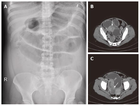
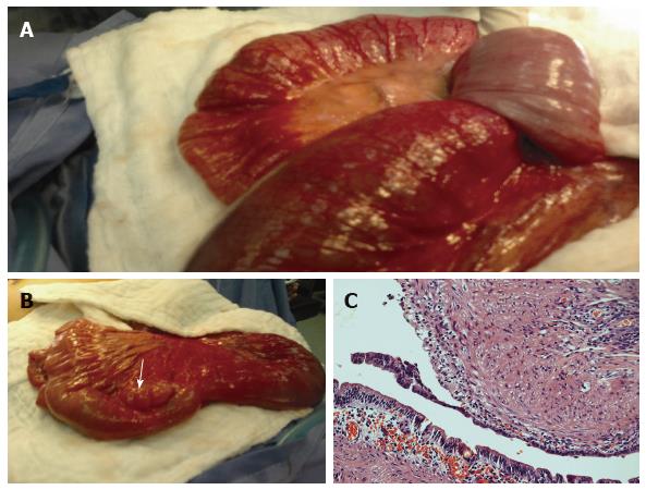
A 32-year-old woman presented to the emergency department (ED) with diffuse intermittent abdominal pain, vomiting, and constipation for 3 d associated with progressive abdominal distention. On examination, she was dehydrated, with a blood pressure of 100/55 mmHg, pulse rate of 110 beat/min, temperature of 37.6 °C. The abdomen was diffusely distended with mild diffuse discomfort and exaggerated bowel sounds. Digital and proctoscopic examinations were normal with empty rectum. Immediate resuscitation in the ED was started with intravenous infusion of crystalloid fluids through large bore intravenous cannulae. A nasogastric tube was inserted which drained large amount of greenish fluids. A Foley catheter was inserted, with drainage of concentrated, minimal amounts of urine. Laboratory values revealed a white blood cell count of 14.6 × 10⁹/L [normal range (NR) = 4.3-10.8], hemoglobin 14.4 g/dL (NR = 14-18), urea 8.5 mmol/L (NR = 3.6-7.1), creatinine 129 mmol/L (NR < 133 mmol/L), and normal coagulation profile. Plain abdominal X-ray revealed dilated small bowel loops (Figure 1). Abdominal computed tomography (CT) showed grossly dilated small bowel loops with “sausage” and “doughnut” signs of small bowel intussusception. She underwent exploratory laparotomy with a provisional diagnosis of small bowel obstruction due to intussusception. Abdominal exploration revealed massively dilated small bowel proximal to ileoileal intussusception. After manual reduction of the intussusception, segmental ileal resection with an end to end anastomosis was performed. Exploration of the resected segment showed the leading point of intussusception was a cystic lesion measuring 4 cm in diameter which was located adjacent to the mesenteric side of the ileum (Figure 2). The histopathology showed an ileal duplication cyst lined by enteric mucosa with distinct muscular layer in the cyst wall. The patient was discharged from the hospital on 7th day postoperatively with an uneventful postoperative course.
DAT has been defined by Ladd et al[4] as a tubular or spherical shaped anomaly that is adherent or attached to and shared the identical phenotypic characteristics with the normal gastrointestinal tract (GIT). It is a rare congenital malformation which can occur anywhere in the GIT from the mouth to the anus, with few cases reported in extraintestinal spaces such as oropharynx, retroperitoneum and spines[5]. It is a separate entity from GIT with their own lumen, which can occur either on the mesenteric or the contralateral side[6]. As the mucosa of the DAT is functional and separate from the intestine, they can expand as their mucosa secret fluids and become symptomatic[6].
The most widely accepted theory for the DAT formation is that the duplication of GIT occurs during embryological development due to a pinching off of diverticulum[5]. The majority of cases have been reported in infants and childhood, however, rarely it has been reported in adults as well[7]. The most commonly reported clinical manifestation of DAT in adult is small bowel obstruction but rarely it may serve as a lead point for intussusception of small bowel, GIT bleeding or iron deficiency anemia due to chronic blood loss[3,5,6]. The etiology of GIT bleeding is varied from abrasion, irritation, or ulceration of mucosal layer within the duplication cyst, or it may arise due to either a disruption of the common blood supply or malignancy which may results in gradual or brisk blood loss[5].
The adult patient reported here presented with a classical symptoms and signs of small bowel obstruction. Intussusception in adults is account for about 5% of all intussusceptions and the underlying cause of only 1% of bowel obstruction[8]. However, a definite underlying cause is present in about 90% of intussusception in adults[9]. A wide variety of ileal lesions may be responsible for ileoileal intussusception. Benign tumors including inflammatory fibroid polyp, hamartomatous polyp, lipoma, as well as malignant tumors such as ileal cancer and lymphoma and Meckel’s diverticulum have all been described as lead points for ileoileal intussusception[3]. In our patient, the lead point was DAT in the mesenteric side of the mid-part of the ileum. Both DAT and intussusception are unusual pathological entities in adult patients, and it is very rare to have the combination of both events in one patient[3]. Although it has been reported in infants and childhood[10,11], to the best of our knowledge, this is the second adult case of DAT identified as a lead point for ileoileal intussusception. Ultrasonography in intussusception may show a “doughnut” configuration on transverse view, while on longitudinal view it may show “pseudokidney” configuration. The detection of excess fluid should raise the suspicion of a lead point lesion[7].
Abdominal CT scan is the most sensitive diagnostic tool for the imaging of intussusceptions. It reveals a pathognomonic appearance of soft tissue mass consisting of the central intussusceptum and the outer intussuscipiens. It appears as “sausage-shaped”, when the CT scan beam is parallel to the intussusception, and it appears as a “target” when the beam is perpendicular to its axis[12]. Clinical and radiological signs of DAT may mimic those of intussusception. Hence, the diagnosis can generally be established only by surgical evaluation[13]. In the present case, CT scan revealed grossly distended proximal small bowel, and both sausage-shape and target signs with diagnosis of small bowel obstruction due to intussusception. However, the DAT was not clear in CT scan as a lead point for intussusception. The differential diagnosis of DAT in association with ileoileal intussusception primarily includes lymphangioma, omental cyst, mesenteric lymphoma, fluid encapsulated in the mesentery during intussusception, Meckel’s diverticulum and intramural neoplasm[3]. A correct and timely diagnosis is necessary not only to resect the underlying lesion that serve as the lead point for intussusception but also to avoid the serious complications of bowel infarction and perforation secondary to obstruction. Resection of the affected bowel and the DAT is recommended to relief the bowel obstruction and to avoid the patient further complications[7]. This patient underwent exploratory laparotomy with presumed diagnosis of complete small bowel obstruction due to intussusception. Abdominal exploration revealed intussusception in the ileum due to cystic lesion located in the mesenteric side of the bowel. The affected bowel was resected with the cystic lesion and end to end bowel anastomosis was established.
In conclusion, a rare case of ileoileal intussusception due to DAT in a young adult woman is reported herein. This case highlights the DAT as an uncommon cause of adult ileoileal intussusception. DAT should be considered in the differential diagnosis of intussusception in adults particularly when the leading point is a cystic lesion. Treatment of choice is resection of the lead point with the affected small intestine and end to end anastomoses.
A 32-year-old woman presented to the emergency department with diffuse intermittent abdominal pain, vomiting, and constipation for 3 d associated with progressive abdominal distention.
The abdomen was diffusely distended with mild diffuse discomfort and exaggerated bowel sounds.
The clinical condition could be small bowel obstruction or large bowel obstruction.
Leukocytic count was high.
Abdominal computed tomography showed grossly dilated small bowel loops with “sausage” and “doughnut” signs of small bowel intussusception.
Exploratory laparotomy, resection of ileoileal intussusception and the leading point was cystic lesion measuring 4 cm in diameter which was located adjacent to the mesenteric side of the ileum.
Enteric duplication cyst as a leading point of intussusception.
Enteric duplication cyst should be considered in the differential diagnosis of intussusception in adults particularly when the leading point is a cystic lesion.
The author describes a rare case of ileoileal intussusception due to enteric duplication cyst in adult patient.
P- Reviewer: Cui J, Klinge U, Li W S- Editor: Qiu S L- Editor: A E- Editor: Wu HL
| 1. | Fiorani C, Scaramuzzo R, Lazzaro A, Biancone L, Palmieri G, Gaspari AL, Sica G. Intestinal duplication in adulthood: A rare entity, difficult to diagnose. World J Gastrointest Surg. 2011;3:128-130. [RCA] [PubMed] [DOI] [Full Text] [Full Text (PDF)] [Cited by in CrossRef: 16] [Cited by in RCA: 24] [Article Influence: 1.7] [Reference Citation Analysis (0)] |
| 2. | Al-Sarem SA, Al-Shawi JS. Ileal duplication in adults. Saudi Med J. 2007;28:1734-1736. [PubMed] |
| 3. | Kim HS, Sung JY, Park WS, Kim YW. An ileal duplication cyst manifested as an ileocolic intussusception in an adult. Turk J Gastroenterol. 2014;25 Suppl 1:196-198. [RCA] [PubMed] [DOI] [Full Text] [Cited by in Crossref: 8] [Cited by in RCA: 9] [Article Influence: 0.9] [Reference Citation Analysis (0)] |
| 4. | Ladd AP, Grosfeld JL. Gastrointestinal tumors in children and adolescents. Semin Pediatr Surg. 2006;15:37-47. [RCA] [PubMed] [DOI] [Full Text] [Cited by in Crossref: 88] [Cited by in RCA: 66] [Article Influence: 3.5] [Reference Citation Analysis (0)] |
| 5. | Tamvakopoulos GS, Sams V, Preston P, Stebbings WS. Iron-deficiency anaemia caused by an enterolith-filled jejunal duplication cyst. Ann R Coll Surg Engl. 2004;86:W49-W51. [RCA] [PubMed] [DOI] [Full Text] [Cited by in Crossref: 11] [Cited by in RCA: 14] [Article Influence: 0.7] [Reference Citation Analysis (0)] |
| 6. | Kim SK, Lim HK, Lee SJ, Park CK. Completely isolated enteric duplication cyst: case report. Abdom Imaging. 2003;28:12-14. [RCA] [PubMed] [DOI] [Full Text] [Cited by in Crossref: 32] [Cited by in RCA: 40] [Article Influence: 1.8] [Reference Citation Analysis (0)] |
| 7. | Shah N, Lizardo-Escaño T, Shaaban H, Dhadham G, Karki A, Spira R. Enterogenous cyst of the small bowel causing intussusception in an adult: Case report and review of literature. J Nat Sci Biol Med. 2015;6:208-210. [RCA] [PubMed] [DOI] [Full Text] [Full Text (PDF)] [Cited by in Crossref: 2] [Cited by in RCA: 3] [Article Influence: 0.3] [Reference Citation Analysis (0)] |
| 9. | Agha FP. Intussusception in adults. AJR Am J Roentgenol. 1986;146:527-531. [RCA] [PubMed] [DOI] [Full Text] [Cited by in Crossref: 209] [Cited by in RCA: 168] [Article Influence: 4.3] [Reference Citation Analysis (0)] |
| 10. | Dias AR, Lopes RI, do Couto RC, Bonafe WW, D’Angelo L, Salvestro ML. Ileal duplication causing recurrent intussusception. J Surg Educ. 2007;64:51-53. [RCA] [PubMed] [DOI] [Full Text] [Cited by in Crossref: 11] [Cited by in RCA: 13] [Article Influence: 0.7] [Reference Citation Analysis (0)] |
| 11. | Maazoun K, Mekki M, Sahnoun L, Hafsa S, Ben Brahim M, Belghith M, Zakhama A, Jouini R, Golli M, Krichene I. Intussusception owing to pathologic lead points in children: report of 27 cases. Arch Pediatr. 2007;14:4-9. [RCA] [PubMed] [DOI] [Full Text] [Cited by in Crossref: 7] [Cited by in RCA: 8] [Article Influence: 0.4] [Reference Citation Analysis (0)] |
| 12. | Gayer G, Zissin R, Apter S, Papa M, Hertz M. Pictorial review: adult intussusception--a CT diagnosis. Br J Radiol. 2002;75:185-190. [RCA] [PubMed] [DOI] [Full Text] [Cited by in Crossref: 148] [Cited by in RCA: 163] [Article Influence: 7.1] [Reference Citation Analysis (0)] |
| 13. | Lahoti HN, Singh RV. Ileo-cecal duplication cyst masquerading as intussusception. Journal of case reports. 2013;3:410-412. [RCA] [DOI] [Full Text] [Cited by in Crossref: 2] [Cited by in RCA: 1] [Article Influence: 0.1] [Reference Citation Analysis (0)] |


