Published online Sep 27, 2013. doi: 10.4240/wjgs.v5.i9.256
Revised: September 9, 2013
Accepted: September 18, 2013
Published online: September 27, 2013
Processing time: 91 Days and 22.1 Hours
An 85-year-old woman presented with sudden onset of generalised abdominal pain and absolute constipation for 4 d. On examination she had a distended abdomen. Plain abdominal radiograph revealed a gas filled viscous within the left upper quadrant. Subsequent computed tomography suggested caecal volvulus herniated through a left diaphragmatic hernia. The patient underwent reduction of the internal hernia, right hemicolectomy and mesh repair of the diaphragmatic hernia. Postoperative recovery was uneventful. Histology revealed a Dukes’ A colonic cancer within the caecum. Herniation of caecal volvulus through a diaphragmatic hernia is a very rare condition and may have been precipitated by the colonic tumour.
Core tip: In patients presenting with acute bowel obstruction rare causes such as caecal volvulus must be included in the differential diagnosis. Herniation of caecal volvulus through a diaphragmatic hernia is a very rare condition and may have been precipitated by the colonic tumour.
- Citation: Bhogal RH, Maleki K, Patel R. Colonic tumour precipitating caecal volvulus within a diaphragmatic hernia. World J Gastrointest Surg 2013; 5(9): 256-258
- URL: https://www.wjgnet.com/1948-9366/full/v5/i9/256.htm
- DOI: https://dx.doi.org/10.4240/wjgs.v5.i9.256
Internal hernias through a diaphragmatic hernia are extremely rare, accounting for only < 1% of all hernias. Diaphragmatic herniae are generally thought to be congenital but other aetiological factors such as trauma have been suggested[1]. The true incidence of diaphragmatic hernia is not known[2]. Furthermore symptoms and/or herniation through such hernias are very rare[1].
Intestinal obstruction due to internal herniation of the caecum in the form of caecal volvulus has not been reported in the literature previously. Caecal volvulus is defined as the axial rotation of the caecum accompanied by a twisting of the mesentery and of its vessels[3]. The classical clinical presentation is that of symptoms of an intestinal obstruction[4]. We report a case of caecal volvulus herniated through a left diaphragmatic hernia that may have been precipitated by a colonic tumour. Prompt surgical intervention following radiological imaging ensured the patient had an excellent post-operative outcome.
An 85-year-old female was admitted with a 4-d history of sudden onset abdominal pain associated with absolute constipation. She had no relevant past medical history and had had no previous abdominal surgery. On examination she had tender but soft abdomen with no signs of peritoneal irritation. The abdomen was markedly distended. There was also hyperactive bowel sound on auscultation. Digital rectal examination demonstrated soft stool in rectum.
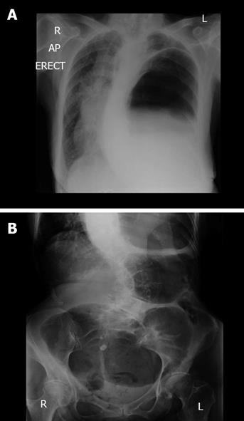
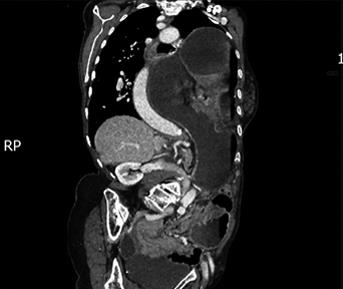
Full blood count, urea and electrolytes, liver function tests, and C-reactive protein were all within normal limits. Erect chest radiograph revealed a collapsed left lung and elevation of the left hemidiaphragm with a large viscous identified under the left hemidiaphragm in keeping with volvulus of the large bowel (Figure 1A). Plain abdominal radiograph revealed a gas filled viscous identified within the left upper quadrant in keeping with volvulus of the caecum (Figure 1B). A computed tomography scan with intravenous contrast was performed subsequently which demonstrated that the caecum had herniated through the diaphragmatic hernia and was causing intestinal obstruction (Figure 2). There were no features suggestive of malignancy.
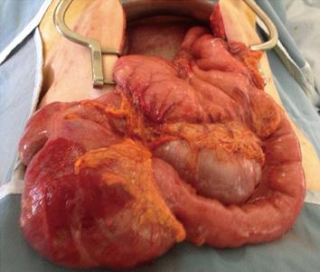
The patient received initial management with intravenous fluid, nasogastric tube drainage, and anti-emetic and analgesic medications. After fluid resuscitation, the patient underwent an emergent laparotomy. A mobile caecum and ascending colon with a long, unfixed mesentery was herniated through the diaphragmatic hernia. The caecum was twisted 270° inside the hernial sac and had developed tears in the outer serous membrane of the bowel termed serosal tears. Furthermore the caecum around the serosal tears appeared congested and ischaemic (Figure 3). A right hemicolectomy with a stapled ileo-transverse colonic anastomosis was performed. The diaphragmatic hernia was repaired with a mesh to prevent further herniation. Specifically the hernia sac was excised and the edges of the hernial defect were freshened. A composite mesh was used for the hernial repair. The polypropylene surface of the mesh was sutured into contact with the diaphragm and the polyglactin surface was left exposed to the peritoneal cavity. Histological analysis of the resected bowel revealed a sessile polyp in the ascending colon measuring 38 mm × 32 mm that was 22 mm from the proximal resection margin. This polyp was a well to moderately differentiated adenocarcinoma arising in a tubulovillous adenoma (pT2, pN0, pMx; Dukes’ A). Histology confirmed the adenoma was infiltrating the muscularis mucosae with the cells displaying an irregular tubular structure. There was no evidence of lymphovascular invasion. The patient’s postoperative recovery was uneventful and she was discharged after 13 wk.
Diaphragmatic hernias resulting from the developmental failure of posterolateral diaphragmatic foramina to fuse properly and was originally described by Salaçin et al[5]. Diaphragmatic hernias typically vary in size, are predominantly left sided, can present at any age and are usually found incidentally[5-7]. Approximately 100 cases of Bochdalek’s hernias in asymptomatic adults have been reported in the literature. The true prevalence of Bochdalek’s hernia remains unknown, with estimates ranging from 1 in 2000-7000[5,6]. Putative causes for late-presenting diaphragmatic hernias include congenital herniation, trauma, physical exertion, pregnancy, sneezing or coughing[5]. The size of the hernia seen on cross-sectional imaging does not necessarily correspond to the size of the diaphragmatic defect, which may be substantially larger[8].
Left-sided diaphragmatic hernias may contain fat, retroperitoneal structures, or intraperitoneal contents, although the latter two conditions are rare[8]. Colon-containing diaphragmatic hernias are exceedingly rare and do usually occur through left-sided defects[9]. The reported case is the first to report a caecal volvulus within a diaphragmatic hernia. Caecal volvulus occurs as a result of axial rotation of the caecum about its mesentery. The ischaemia and serosal tears observed in the reported patient are likely due to venous congestion of the caecum due to the twisted mesentery[10]. Other authors have also reported that intestinal volvulus causes intestinal ischaemia[11]. Previous reports in the literature suggest that tumours may precipitate volvulus[12,13]. The putative mechanism suggests that high peristaltic bowel centred on the tumour cause a loop of bowel to descend inferiorly. This displaces empty small bowel loops upwards, initiating rotation of the mesentery and causing volvulus[13].
Martin et al[14] described the first open repair of a diaphragmatic hernia. Although laparoscopic repair of a diaphragmatic hernia has been described this approach is probably best reserved for the elective setting[15]. In our case after the standard right hemicolectomy was performed for the caecal volvulus the diaphragmatic hernia required repair. Consensus on whether synthetic mesh or primary closure produce the safest and most durable repair for diaphragmatic hernia has yet to be agreed[16]. The placement of synthetic mesh repair in close proximity to the esophagus runs the risk of erosion[17]. Synthetic mesh was chosen for the large diaphragmatic defect as in our case. Ethicon Vypro mesh was used for its dual surface properties. It has a fascial side, which induces tissue in-growth and thus results in better tissue fixation. On the other hand, on its peritoneal side it has a low-porosity smooth visceral surface that minimizes visceral adhesion.
In summary, precise radiological evaluation and prompt surgical repair results in good outcome in patients presenting with bowel obstruction secondary to a diaphragmatic hernia.
P- Reviewers Dhawan DK, Kir G, Shida D S- Editor Gou SX L- Editor A E- Editor Wang CH
| 1. | Brown SR, Horton JD, Trivette E, Hofmann LJ, Johnson JM. Bochdalek hernia in the adult: demographics, presentation, and surgical management. Hernia. 2011;15:23-30. [RCA] [PubMed] [DOI] [Full Text] [Cited by in Crossref: 66] [Cited by in RCA: 83] [Article Influence: 5.5] [Reference Citation Analysis (1)] |
| 2. | Schumacher L, Gilbert S. Congenital diaphragmatic hernia in the adult. Thorac Surg Clin. 2009;19:469-472. [RCA] [PubMed] [DOI] [Full Text] [Cited by in Crossref: 35] [Cited by in RCA: 28] [Article Influence: 1.9] [Reference Citation Analysis (0)] |
| 3. | Ruiz-Tovar J, Calero García P, Morales Castiñeiras V, Martínez Molina E. Caecal volvulus: presentation of 18 cases and review of literature. Cir Esp. 2009;85:110-113. [RCA] [PubMed] [DOI] [Full Text] [Cited by in Crossref: 11] [Cited by in RCA: 10] [Article Influence: 0.6] [Reference Citation Analysis (0)] |
| 4. | Consorti ET, Liu TH. Diagnosis and treatment of caecal volvulus. Postgrad Med J. 2005;81:772-776. [RCA] [PubMed] [DOI] [Full Text] [Cited by in Crossref: 100] [Cited by in RCA: 107] [Article Influence: 5.6] [Reference Citation Analysis (0)] |
| 5. | Salaçin S, Alper B, Cekin N, Gülmen MK. Bochdalek hernia in adulthood: a review and an autopsy case report. J Forensic Sci. 1994;39:1112-1116. [PubMed] |
| 6. | Nitecki S, Bar-Maor JA. Late presentation of Bochdalek hernia: our experience and review of the literature. Isr J Med Sci. 1992;28:711-714. [PubMed] |
| 7. | Gale ME. Bochdalek hernia: prevalence and CT characteristics. Radiology. 1985;156:449-452. [PubMed] |
| 8. | Wilbur AC, Gorodetsky A, Hibbeln JF. Imaging findings of adult Bochdalek hernias. Clin Imaging. 1994;18:224-229. [RCA] [PubMed] [DOI] [Full Text] [Cited by in Crossref: 28] [Cited by in RCA: 32] [Article Influence: 1.0] [Reference Citation Analysis (0)] |
| 9. | Bétrémieux P, Dabadie A, Chapuis M, Pladys P, Tréguier C, Frémond B, Lefrancois C. Late presenting Bochdalek hernia containing colon: misdiagnosis risk. Eur J Pediatr Surg. 1995;5:113-115. [RCA] [PubMed] [DOI] [Full Text] [Cited by in Crossref: 28] [Cited by in RCA: 29] [Article Influence: 1.0] [Reference Citation Analysis (0)] |
| 10. | Pousada L. Cecal bascule: an overlooked diagnosis in the elderly. J Am Geriatr Soc. 1992;40:65-67. [PubMed] |
| 11. | Vokurka J, Olejnik J, Jedlicka V, Vesely M, Ciernik J, Paseka T. Acute mesenteric ischemia. Hepatogastroenterology. 2008;55:1349-1352. [PubMed] |
| 12. | Cengız F, Sun MA, Esen ÖS, Erkan N. Gastrointestinal stromal tumor of Meckel’s diverticulum: a rare cause of intestinal volvulus. Turk J Gastroenterol. 2012;23:410-412. [PubMed] |
| 13. | Leong C. Ileal volvulus and its association with carcinoid tumours. Australas Med J. 2012;5:326-328. [RCA] [PubMed] [DOI] [Full Text] [Cited by in Crossref: 1] [Cited by in RCA: 2] [Article Influence: 0.2] [Reference Citation Analysis (0)] |
| 14. | Martin I, O’Rourke N, Gotley D, Smithers M. Laparoscopy in the management of diaphragmatic rupture due to blunt trauma. Aust N Z J Surg. 1998;68:584-586. [RCA] [PubMed] [DOI] [Full Text] [Cited by in Crossref: 25] [Cited by in RCA: 27] [Article Influence: 1.0] [Reference Citation Analysis (0)] |
| 15. | Frantzides CT, Carlson MA. Laparoscopic repair of a penetrating injury to the diaphragm: a case report. J Laparoendosc Surg. 1994;4:153-156. [RCA] [PubMed] [DOI] [Full Text] [Cited by in Crossref: 23] [Cited by in RCA: 24] [Article Influence: 0.8] [Reference Citation Analysis (0)] |
| 16. | Contini S, Dalla Valle R, Bonati L, Zinicola R. Laparoscopic repair of a Morgagni hernia: report of a case and review of the literature. J Laparoendosc Adv Surg Tech A. 1999;9:93-99. [RCA] [PubMed] [DOI] [Full Text] [Cited by in Crossref: 38] [Cited by in RCA: 37] [Article Influence: 1.4] [Reference Citation Analysis (0)] |
| 17. | Matthews BD, Bui H, Harold KL, Kercher KW, Adrales G, Park A, Sing RF, Heniford BT. Laparoscopic repair of traumatic diaphragmatic injuries. Surg Endosc. 2003;17:254-258. [RCA] [PubMed] [DOI] [Full Text] [Cited by in Crossref: 84] [Cited by in RCA: 87] [Article Influence: 4.0] [Reference Citation Analysis (0)] |


